
Anti-AMPK alpha 1 PRKAA1 Rabbit Monoclonal Antibody, Clone: [Clone: CDD-16]
Artikelnummer:
BOB-M00994
- Bilder (9)
| Artikelname: | Anti-AMPK alpha 1 PRKAA1 Rabbit Monoclonal Antibody, Clone: [Clone: CDD-16] |
| Artikelnummer: | BOB-M00994 |
| Hersteller Artikelnummer: | M00994 |
| Alternativnummer: | BOB-M00994-100UL |
| Hersteller: | Boster Bio |
| Wirt: | Rabbit |
| Kategorie: | Antikörper |
| Applikation: | FC, ICC, IF, IHC, IP, WB |
| Spezies Reaktivität: | Human, Mouse, Rat |
| Immunogen: | A synthesized peptide derived from human AMPK alpha 1 |
| Alternative Synonym: | AMPK, AMPK alpha, AMPK alpha 1, AMPK subunit alpha 1, AMPK1, AMPKa1, PRKAA1 |
| Boster Bio Anti-AMPK alpha 1 PRKAA1 Rabbit Monoclonal Antibody catalog M00994. Tested in WB, IHC, ICC/IF, IP, Flow Cytometry applications. This antibody reacts with Human, Mouse, Rat. |
| Klonalität: | Monoclonal |
| Konzentration: | 0.5mg/ml |
| Klon-Bezeichnung: | [Clone: CDD-16] |
| Molekulargewicht: | Observed Molecular Weight: 64 kDa. Calculated Molecular Weight: 64009 MW |
| NCBI: | 5562 |
| UniProt: | Q13131 |
| Puffer: | Rabbit IgG in stabilizing components, phosphate buffered saline, pH 7.4, 150mM NaCl, 0.02% sodium azide and 50% glycerol. *This antibody is supplied in a stabilized formulation. Compatibility with conjugation reactions depends on the chemistry of the con |
| Reinheit: | Affinity-chromatography |
| Formulierung: | Liquid |
| Target-Kategorie: | 5-AMP-activated protein kinase catalytic subunit alpha-1 |
| Application Verdünnung: | WB 1:500-2000IHC 1:50-200ICC/IF 1:50-200IP 1:20FC 1:20 |
|
|
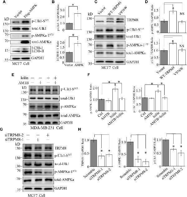
Involvement of the AMPK-ULK1-LC3 signaling cascade in TRPM8-stimulated autophagy. (A, B) The construct for Flag-AMPK expression was transiently transfected into MCF7 cells. After 48h of transfection, the AMPK-ULK1-LC3 signaling cascade-related proteins were detected by WB (N = 3). (C, D) MCF7 cells were transiently transfected with wild type TRPM8, mutant V976W, or control vector. After 48h of transfection, protein lysates were used for WB analysis (N = 3). (E, F) MDA-MB-231 cells treated with 2 µM icilin, 0.5 µM AMTB, or their combination for 48h were extracted for WB analysis (N = 3). (G, H) MCF7 cells were transfected with siRNA against human TRPM8, siRNA against TRPM8 (siTRPM8-1 and siTRPM8-2) successfully knocked down TRPM8 expression compared with that in the control scramble siRNA samples according to WB analysis using an anti-TRPM8 antibody. The AMPK-ULK1-LC3 signaling cascade-related proteins detected by WB analysis using the indicated antibodies (N = 3). N represents the number of replicate experiments. *P < 0.05, NS, not significant.Index in PubMed under a CC BY license. PMID: 33344232 |
|
|
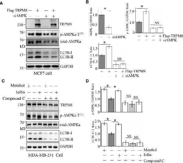
Influence of AMPK impairment on the stimulatory effect of TRPM8 on autophagy. (A, B) MCF7 cells were transiently transfected with siRNA against human AMPK and a Flag-TRPM8 construct. After 48h of transfection, protein lysates were extracted for WB analysis to determine the effect of TRPM8 overexpression on basal autophagy in the presence of AMPK knockdown (N = 3). (C, D) WB analysis of cell lysates of MDA-MB-231 cells treatment with 2 µM icilin, 10 µM menthol, or a combination of 10 M compound C for 48h (N = 3). N represents the number of replicate experiments. *P < 0.05, NS, not significant.Index in PubMed under a CC BY license. PMID: 33344232 |
|
|
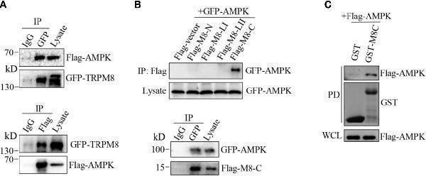
AMPK interacts with TRPM8. (A, B) Co-IP analysis. (A) Constructs for GFP-TRPM8 and Flag-AMPK expression were transiently transfected into MCF7 cells. After 48h of transfection, protein lysates were immunoprecipitated with an anti-GFP antibody and assayed by immunobl |
|
|
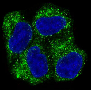
Immunofluorescent analysis of Hela cells, using AMPK alpha 1 Antibody . |
|
|
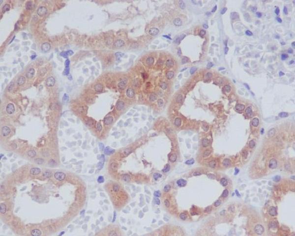
Immunohistochemical analysis of paraffin-embedded human kidney, using AMPK alpha 1 Antibody. |
|
|
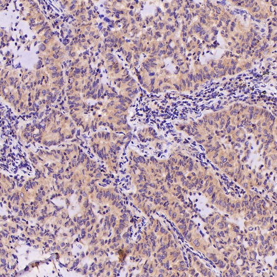
Immunohistochemical analysis of paraffin-embedded Human lung adenocarcinoma, using the Antibody at 1:500 dilution. |
|
|
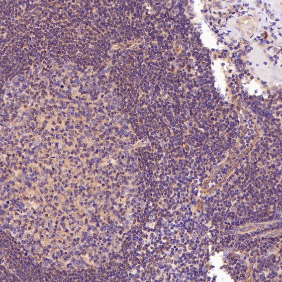
Immunohistochemical analysis of paraffin-embedded Human tonsil, using the Antibody at 1:500 dilution. |
|
|
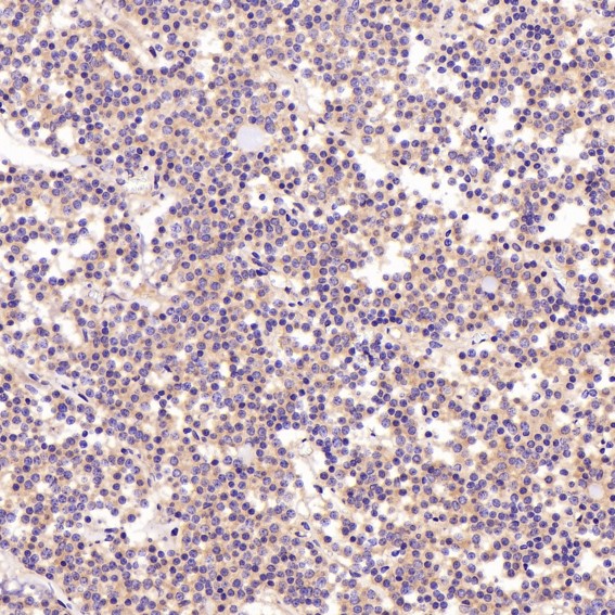
Immunohistochemical analysis of paraffin-embedded Human pituitary tumor, using the Antibody at 1:500 dilution. |
|
|
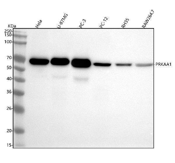
Western blot analysis of AMPK alpha 1 using anti-AMPK alpha 1 antibody (M00994). Electrophoresis was performed on a 5-20% SDS-PAGE gel at 70V (Stacking gel) / 90V (Resolving gel) for 2-3 hours. The sample well of each lane was loaded with 30 ug of sample under reducing conditions. Lane 1: human Hela whole cell lysates,Lane 2: human U-87MG whole cell lysates,Lane 3: human PC-3 whole cell lysates,Lane 4: rat PC-12 whole cell lysates,Lane 5: rat RH35 whole cell lysates,Lane 6: mouse RAW264.7 whole cell lysates.After electrophoresis, proteins were transferred to a nitrocellulose membrane at 150 mA for 50-90 minutes. Blocked the membrane with 5% non-fat milk/TBS for 1.5 hour at RT. The membrane was incubated with rabbit anti-AMPK alpha 1 antigen affinity purified monoclonal antibody (Catalog M00994) at 1:500 overnight at 4C, then washed with TBS-0.1%Tween 3 times with 5 minutes each and probed with a goat anti-rabbit IgG-HRP secondary antibody at a dilution of 1:500 for 1.5 hour at RT. The signal is developed using an Enhanced Chemiluminescent detection (ECL) kit (Catalog EK1002) with Tanon 5200 system. A specific band was detected for AMPK alpha 1 at approximately 64 kDa. The expected band size for AMPK alpha 1 is at 64 kDa. |
Produktgarantie und fachkundiger Support
