
Anti-Calreticulin Rabbit Monoclonal Antibody, Clone: [Clone: CGO-3]
Artikelnummer:
BOB-M00894
- Bilder (9)
| Artikelname: | Anti-Calreticulin Rabbit Monoclonal Antibody, Clone: [Clone: CGO-3] |
| Artikelnummer: | BOB-M00894 |
| Hersteller Artikelnummer: | M00894 |
| Alternativnummer: | BOB-M00894-100UL |
| Hersteller: | Boster Bio |
| Wirt: | Rabbit |
| Kategorie: | Antikörper |
| Applikation: | FC, ICC, IF, IHC, IP, WB |
| Spezies Reaktivität: | Human, Mouse, Rat |
| Immunogen: | A synthesized peptide derived from human Calreticulin - ER Marker |
| Alternative Synonym: | CALR, Calregulin, calreticulin, cC1qR, CRP55, CRT, CRTC, ERp60, grp60, HACBP, RO, SSA |
| Boster Bio Anti-Calreticulin Rabbit Monoclonal Antibody catalog M00894. Tested in WB, IHC, ICC/IF, IP, Flow Cytometry applications. This antibody reacts with Human, Mouse, Rat. |
| Klonalität: | Monoclonal |
| Konzentration: | 0.5mg/ml |
| Klon-Bezeichnung: | [Clone: CGO-3] |
| Molekulargewicht: | Observed Molecular Weight: 60 kDa. Calculated Molecular Weight: 48142 MW |
| NCBI: | 811 |
| UniProt: | P27797 |
| Puffer: | Rabbit IgG in stabilizing components, phosphate buffered saline, pH 7.4, 150mM NaCl, 0.02% sodium azide and 50% glycerol. *This antibody is supplied in a stabilized formulation. Compatibility with conjugation reactions depends on the chemistry of the con |
| Reinheit: | Affinity-chromatography |
| Formulierung: | Liquid |
| Target-Kategorie: | Calreticulin |
| Application Verdünnung: | WB 1:500-2000IHC 1:50-200ICC/IF 1:50-200IP 1:20FC 1:20 |
|
|
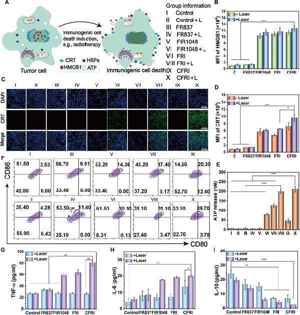
(A) CFRI-mediated ICD and DCs matured invitro. Mechanism scheme of PTT-induced immunogenic death, 1,064-nm laser irradiation (1.0 W/cm 2 ) with or without treatment with DMEM, FR837, FIR1048, FRI, and CFRI (2 µg/ml). (B) The efflux of HMGB1 was quantitatively analyzed by flow cytometry. (C) CLSM images of CRT expression on the 4T1 cell surface after different treatments (scale bar: 100 µm). (D) Mean fluorescence intensity of CRT expression in 4T1 cells after different treatments. (E) The average fluorescence intensity of ATP exposure in 4T1 cells after different treatments. (F) The corresponding quantification (CD80 + CD86 + ) of mature DCs was quantitatively analyzed by flow cytometry. (G) After different treatments by enzyme-linked immunosorbent assay (ELISA), secretion levels of TNF-a in the cell supernatant. (H) After different treatments by ELISA, the secretion level of IL-6 in the cell supernatant. (I) After different treatments by ELISA, the secretion level of IL-10 in the cell supernatant. Information of each group: I, control, II, control + L, III, FR837, IV, FR837 + L, V, FIR1048, VI, FIR1048 + L, VII, FRI, VII, FRI + L, IX, CFRI, X, CFRI + L. Significance is determined using one-way analysis of variance (* P < 0.05, ** P < 0.01, and *** P < 0.001). HSPs, heat shock proteins.Index in PubMed under a CC BY license. PMID: 40040955 |
|
|
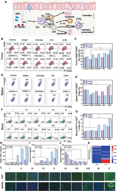
In vivo CFRI-mediated PTT-enhanced immunotherapy elicited an immune response after 16 d of treatment. (A) Schematic diagram of immunity induced by damage-associated molecular patterns (DAMPs). (B and C) Analysis of DC maturity (CD11 + CD80 + CD86 + ) using flow cytometry in the primary tumor of 4T1 tumor-bearing mice. (D and E) Regulatory T cells (Tregs) (CD4 + CD25 + ) in the spleen. (F and G) T cell proliferation in the blood (CD4 + CD8 + ). (H) The content of TNF-alpha in the spleen of 4T1 tumor-bearing mice. (I) The content of IL-6 in the spleen of 4T1 tumor-bearing mice. (J) The content of IL-10 in the |
|
|
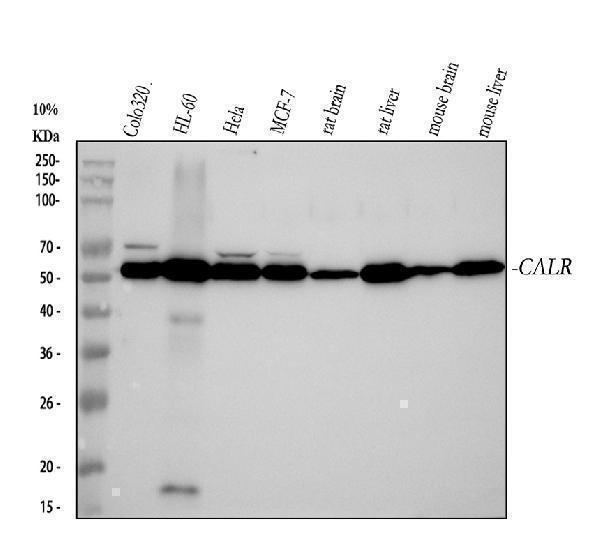
Western blot analysis of Calreticulin using anti-Calreticulin antibody (M00894). Electrophoresis was performed on a 5-20% SDS-PAGE gel at 70V (Stacking gel) / 90V (Resolving gel) for 2-3 hours. The sample well of each lane was loaded with 30 ug of sample under reducing conditions. Lane 1: human COLO320 whole cell lysates,Lane 2: human HL-60 whole cell lysates,Lane 3: human Hela whole cell lysates,Lane 4: human MCF-7 whole cell lysates,Lane 5: rat brain tissue lysates,Lane 6: rat liver tissue lysates,Lane 7: mouse brain tissue lysates,Lane 8: mouse liver tissue lysates.After electrophoresis, proteins were transferred to a nitrocellulose membrane at 150 mA for 50-90 minutes. Blocked the membrane with 5% non-fat milk/TBS for 1.5 hour at RT. The membrane was incubated with rabbit anti-Calreticulin antigen affinity purified monoclonal antibody (Catalog M00894) at 1:500 overnight at 4C, then washed with TBS-0.1%Tween 3 times with 5 minutes each and probed with a goat anti-rabbit IgG-HRP secondary antibody at a dilution of 1:5000 for 1.5 hour at RT. The signal is developed using an Enhanced Chemiluminescent detection (ECL) kit (Catalog EK1002) with Tanon 5200 system. A specific band was detected for Calreticulin at approximately 60 kDa. The expected band size for Calreticulin is at 48 kDa. |
|
|
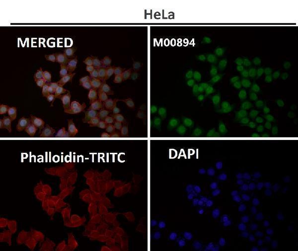
Immunofluorescent analysis using the Antibody at 1:50 dilution. |
|
|
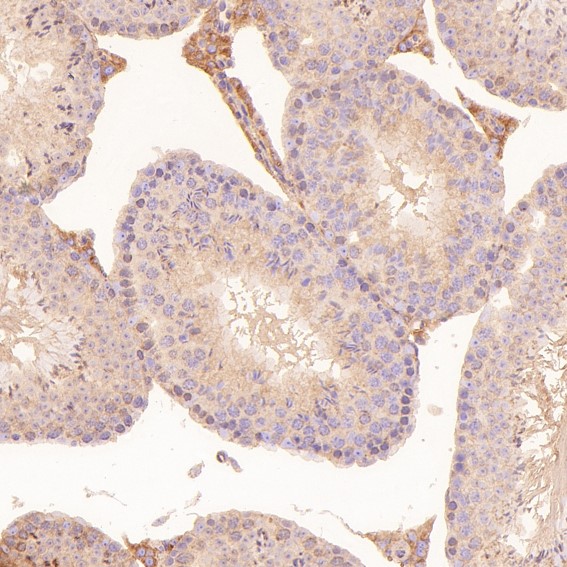
Immunohistochemical analysis of paraffin-embedded Mouse testis, using the Antibody at 1:150 dilution. |
|
|
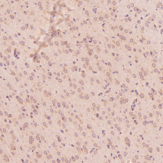
Immunohistochemical analysis of paraffin-embedded Mouse cerebellum, using the Antibody at 1:150 dilution. |
|
|
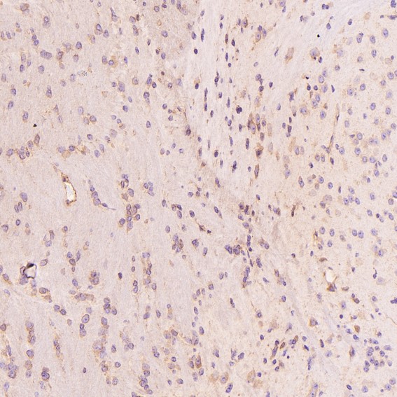
Immunohistochemical analysis of paraffin-embedded Rat cerebral cortex, using the Antibody at 1:150 dilution. |
|
|
Immunohistochemical analysis of paraffin-embedded Human renal cancer, using the Antibody at 1:150 dilution. |
|
|
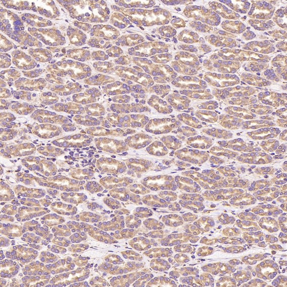
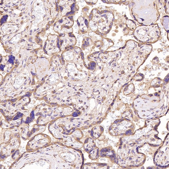
Immunohistochemical analysis of paraffin-embedded Human placenta, using the Antibody at 1:150 dilution. |
Produktgarantie und fachkundiger Support
