PBS (pH 7.3) containing 1% stabilizing protein, 50% glycerol and 0.02% sodium azide.This antibody is supplied in a stabilized formulation. Compatibility with conjugation reactions depends on the chemistry of the conjugation method used. For conjugation me
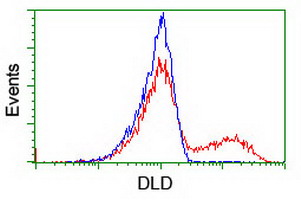
HEK293T cells transfected with either DLD (Myc-DDK-tagged) overexpress plasmid (Red) or empty vector control plasmid (Blue) were immunostained by anti-DLD antibody (M00870)
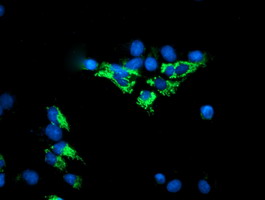
Anti-DLD mouse monoclonal antibody (M00870) immunofluorescent staining of COS7 cells transiently transfected by pCMV6-ENTRY DLD.
Immunohistochemical staining of paraffin-embedded Human pancreas tissue within the normal limits using anti-DLD mouse monoclonal antibody. (Heat-induced epitope retrieval by 10mM citric buffer
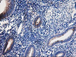
Immunohistochemical staining of paraffin-embedded Human endometrium tissue within the normal limits using anti-DLD mouse monoclonal antibody. (Heat-induced epitope retrieval by 10mM citric buffer
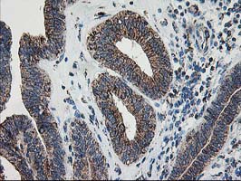
Immunohistochemical staining of paraffin-embedded Adenocarcinoma of Human endometrium tissue using anti-DLD mouse monoclonal antibody. (Heat-induced epitope retrieval by 10mM citric buffer
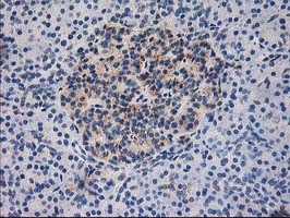
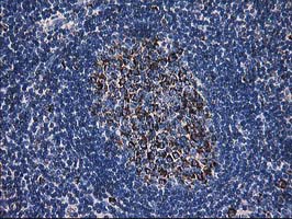
Immunohistochemical staining of paraffin-embedded Human lymph node tissue within the normal limits using anti-DLD mouse monoclonal antibody. (Heat-induced epitope retrieval by 10mM citric buffer
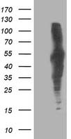
HEK293T cells were transfected with the pCMV6-ENTRY control (Left lane) or pCMV6-ENTRY DLD (Right lane) cDNA for 48 hrs and lysed. Equivalent amounts of cell lysates (5 ug per lane) were separated by SDS-PAGE and immunoblotted with anti-DLD.
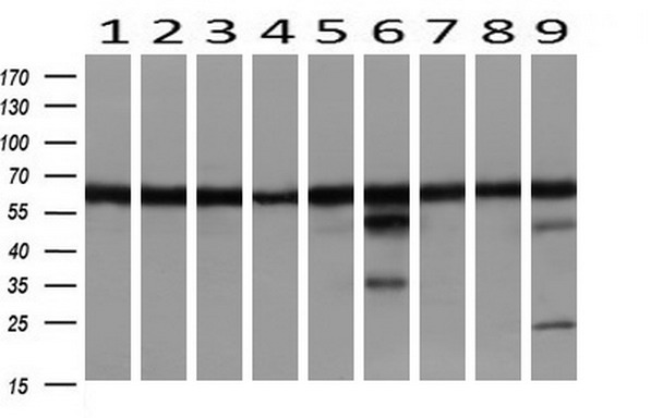
Western blot analysis of extracts (10ug) from 9 Human tissue by using anti-DLD monoclonal antibody at 1:1000 (1: Testis, 2: Omentum, 3: Uterus, 4: Breast, 5: Brain, 6: Liver, 7: Ovary, 8: Thyroid gland, 9: colon).
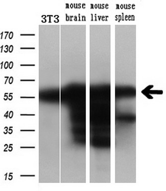
Western blot analysis of extracts (10ug) from a mouse cell line and 3 different mouse tissues by using anti-DLD monoclonal antibody (1:200).
* Mehrwertsteuer und Versandkosten nicht enthalten. Irrtümer und Preisänderungen vorbehalten