DELTA, Delta transcription factor, INO80 complex subunit S, INO80S, NF E1, UCRBP, Yin and yang 1, YIN YANG 1, YY 1, YY1, YY1 transcription factor
Boster Bio Anti-YY1 Rabbit Monoclonal Antibody catalog M00833. Tested in WB, IHC, ICC/IF, IP applications. This antibody reacts with Human, Mouse, Rat.
Rabbit IgG in stabilizing components, phosphate buffered saline, pH 7.4, 150mM NaCl, 0.02% sodium azide and 50% glycerol. *This antibody is supplied in a stabilized formulation. Compatibility with conjugation reactions depends on the chemistry of the con
Reinheit:
Affinity-chromatography
Formulierung:
Liquid
Target-Kategorie:
Transcriptional repressor protein YY1
Application Verdünnung:
WB 1:500-2000IHC 1:50-200ICC/IF 1:50-200IP 1:20
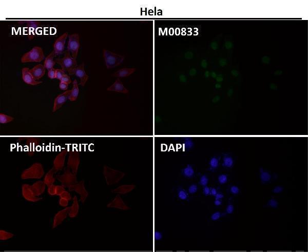
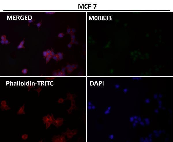
Immunofluorescent analysis using the Antibody at 1:50 dilution.
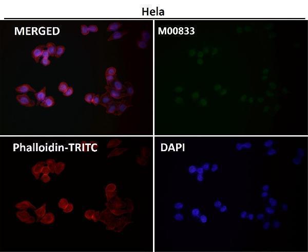
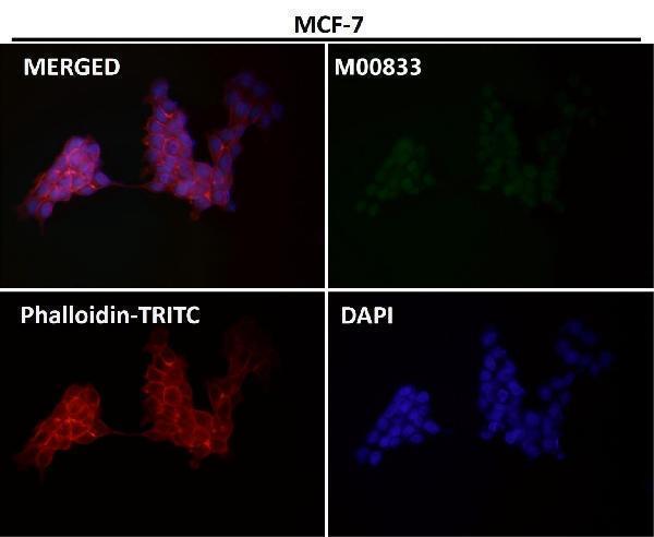
Immunofluorescent analysis using the Antibody at 1:150 dilution.
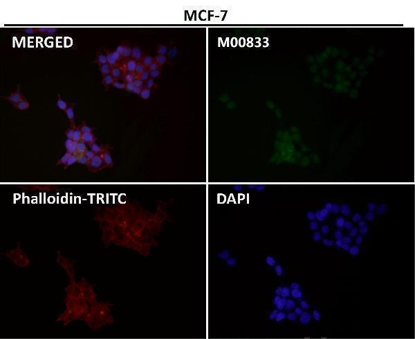
Immunofluorescent analysis using the Antibody at 1:50 dilution.
Immunofluorescent analysis using the Antibody at 1:50 dilution.
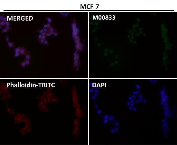
Immunofluorescent analysis using the Antibody at 1:150 dilution.
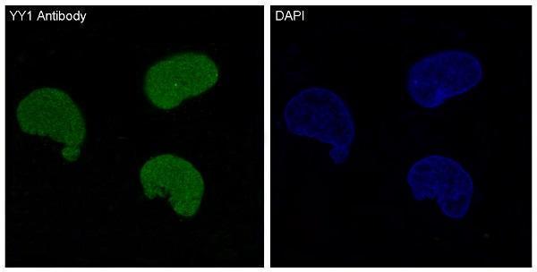
Immunofluorescent analysis using the Antibody at 1:500 dilution.
Immunofluorescent analysis of Hela cells, using YY1 Antibody.
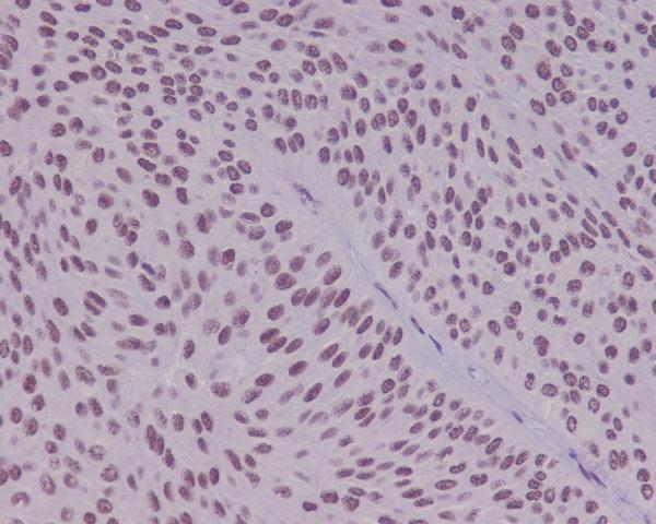
Immunohistochemical analysis of paraffin-embedded human bladder using YY1 Antibody.
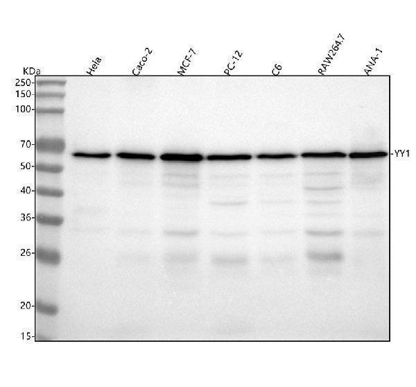
Western blot analysis of YY1 using anti-YY1 antibody (M00833). Electrophoresis was performed on a 5-20% SDS-PAGE gel at 70V (Stacking gel) / 90V (Resolving gel) for 2-3 hours. The sample well of each lane was loaded with 30 ug of sample under reducing conditions. Lane 1: human Hela whole cell lysates,Lane 2: human CACO-2 whole cell lysates,Lane 3: human MCF-7 whole cell lysates,Lane 4: rat PC-12 whole cell lysates,Lane 5: rat C6 whole cell lysates,Lane 6: mouse RAW264.7 whole cell lysates,Lane 7: mouse ANA-1 whole cell lysates.After electrophoresis, proteins were transferred to a nitrocellulose membrane at 150 mA for 50-90 minutes. Blocked the membrane with 5% non-fat milk/TBS for 1.5 hour at RT. The membrane was incubated with rabbit anti-YY1 antigen affinity purified monoclonal antibody (Catalog M00833) at 1:500 overnight at 4C, then washed with TBS-0.1%Tween 3 times with 5 minutes each and probed with a goat anti-rabbit IgG-HRP secondary antibody at a dilution of 1:500 for 1.5 hour at RT. The signal is developed using an Enhanced Chemiluminescent detection (ECL) kit (Catalog EK1002) with Tanon 5200 system. A specific band was detected for YY1 at approximately 68 kDa. The expected band size for YY1 is at 45 kDa.
* Mehrwertsteuer und Versandkosten nicht enthalten. Irrtümer und Preisänderungen vorbehalten