A synthetic peptide corresponding to a sequence at the N-terminus of human EEF2/Elongation factor 2, identical to the related mouse and rat sequences.
Alternative Synonym:
EEF 2, EEF2, EF 2, EF2, Elongation factor 2
Boster Bio Anti-EEF2 Picoband Antibody (monoclonal, 5F5) catalog M00830-2. Tested in Flow Cytometry, IF, IHC, ICC, WB applications. This antibody reacts with Human, Mouse, Rat. The brand Picoband indicates this is a premium antibody that guarantees superior quality, high affinity, and strong signals with minimal background in Western blot applications. Only our best-performing antibodies are designated as Picoband, ensuring unmatched performance.
Klonalität:
Monoclonal
Konzentration:
Adding 0.2 ml of distilled water will yield a concentration of 500 µg/ml.
Each vial contains 4mg Trehalose, 0.9mg NaCl and 0.2mg Na2HPO4.
Reinheit:
Immunogen affinity purified.
Formulierung:
Lyophilized
Target-Kategorie:
Elongation factor 2
Application Verdünnung:
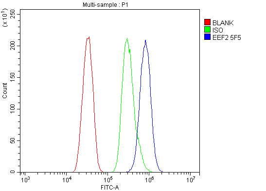
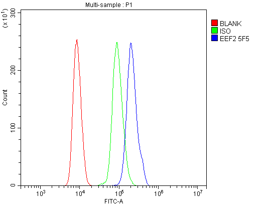
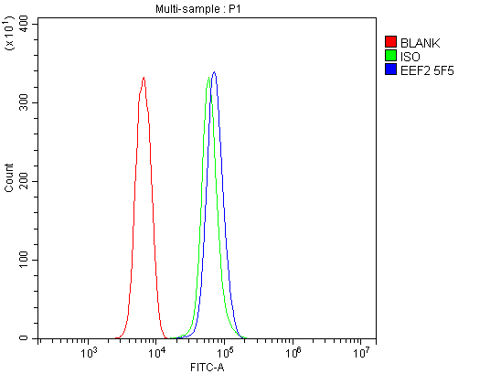
Western blot, 0.25-0.5µg/ml, Human, Mouse, Rat Immunohistochemistry (Paraffin-embedded Section), 2-5µg/ml, Huma Immunocytochemistry/Immunofluorescence, 5µg/ml, Human Flow Cytometry (Fixed), 1-3µg/1x106 cells, Human, Mouse, Rat
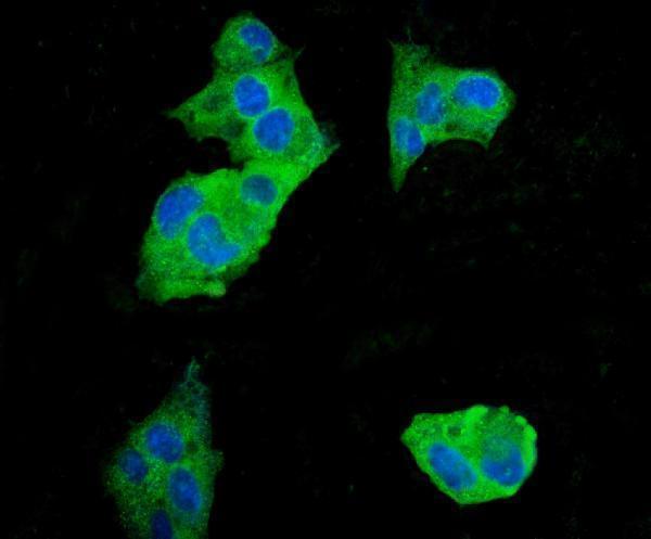
IF analysis of EEF2 using anti-EEF2 antibody (M00830-2). EEF2 was detected in immunocytochemical section of HEPG2 cells. Enzyme antigen retrieval was performed using IHC enzyme antigen retrieval reagent (AR0022) for 15 mins. The cells were blocked with 10% goat se
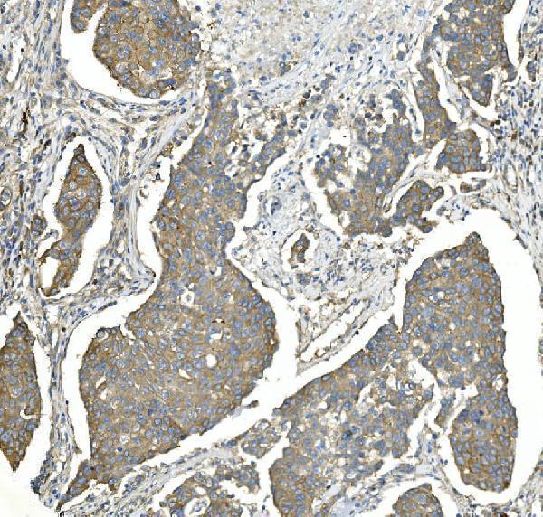
IHC analysis of EEF2 using anti-EEF2 antibody (M00830-2). EEF2 was detected in paraffin-embedded section of human breast cancer tissue. Heat mediated antigen retrieval was performed in EDTA buffer (pH8.0, epitope retrieval solution). The tissue section was blocked with 10% goat serum. The tissue section was then incubated with 2µg/ml mouse anti-EEF2 Antibody (M00830-2) overnight at 4C. Biotinylated goat anti-mouse IgG was used as secondary antibody and incubated for 30 minutes at 37C. The tissue section was developed using Strepavidin-Biotin-Complex (SABC) (Catalog SA1021) with DAB as the chromogen.
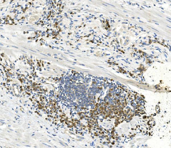
IHC analysis of EEF2 using anti-EEF2 antibody (M00830-2). EEF2 was detected in paraffin-embedded section of human rectal cancer tissue. Heat mediated antigen retrieval was performed in EDTA buffer (pH8.0, epitope retrieval solution). The tissue section was blocked with 10% goat serum. The tissue section was then incubated with 2µg/ml mouse anti-EEF2 Antibody (M00830-2) overnight at 4C. Biotinylated goat anti-mouse IgG was used as secondary antibody and incubated for 30 minutes at 37C. The tissue section was developed using Strepavidin-Biotin-Complex (SABC) (Catalog SA1021) with DAB as the chromogen.
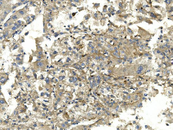
IHC analysis of EEF2 using anti-EEF2 antibody (M00830-2). EEF2 was detected in paraffin-embedded section of human liver cancer tissue. Heat mediated antigen retrieval was performed in EDTA buffer (pH8.0, epitope retrieval solution). The tissue section was blocked with 10% goat serum. The tissue section was then incubated with 2µg/ml mouse anti-EEF2 Antibody (M00830-2) overnight at 4C. Biotinylated goat anti-mouse IgG was used as secondary antibody and incubated for 30 minutes at 37C. The tissue section was developed using Strepavidin-Biotin-Complex (SABC) (Catalog SA1021) with DAB as the chromogen.
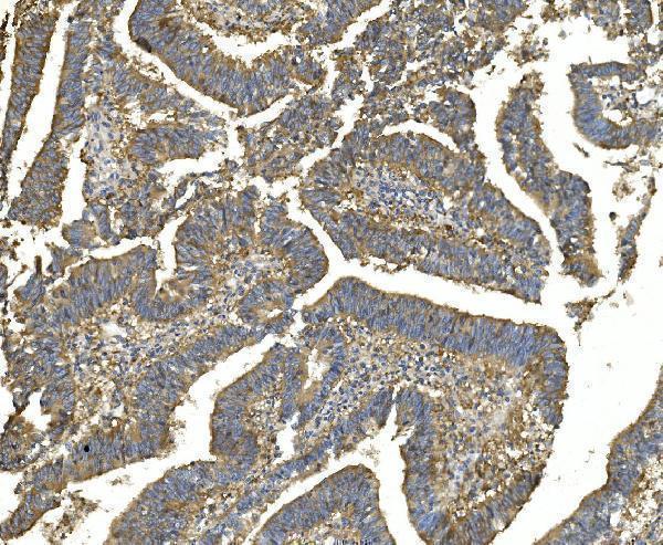
IHC analysis of EEF2 using anti-EEF2 antibody (M00830-2). EEF2 was detected in paraffin-embedded section of human rectal cancer tissue. Heat mediated antigen retrieval was performed in EDTA buffer (pH8.0, epitope retrieval solution). The tissue section was blocked with 10% goat serum. The tissue section was then incubated with 2µg/ml mouse anti-EEF2 Antibody (M00830-2) overnight at 4C. Biotinylated goat anti-mouse IgG was used as secondary antibody and incubated for 30 minutes at 37C. The tissue section was developed using Strepavidin-Biotin-Complex (SABC) (Catalog SA1021) with DAB as the chromogen.
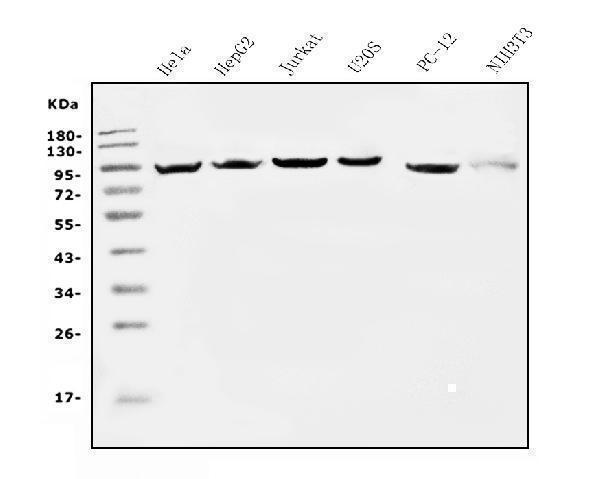
Western blot analysis of EEF2 using anti-EEF2 antibody (M00830-2). Electrophoresis was performed on a 5-20% SDS-PAGE gel at 70V (Stacking gel) / 90V (Resolving gel) for 2-3 hours. The sample well of each lane was loaded with 50ug of sample under reducing conditions. Lane 1: human Hela whole cell lysates, Lane 2: human HEPG2 whole cell lysates, Lane 3: human Jurkat whole cell lysates, Lane 4: human U20S whole cell lysates, Lane 5: rat PC-12 whole cell lysates, Lane 6: mouse NIH/3T3 whole cell lysates. After Electrophoresis, proteins were transferred to a Nitrocellulose membrane at 150mA for 50-90 minutes. Blocked the membrane with 5% Non-fat Milk/ TBS for 1.5 hour at RT. The membrane was incubated with mouse anti-EEF2 antigen affinity purified monoclonal antibody (Catalog M00830-2) at 0.5 µg/mL overnight at 4C, then washed with TBS-0.1%Tween 3 times with 5 minutes each and probed with a goat anti-mouse IgG-HRP secondary antibody at a dilution of 1:10000 for 1.5 hour at RT. The signal is developed using an Enhanced Chemiluminescent detection (ECL) kit (Catalog EK1001) with Tanon 5200 system. A specific band was detected for EEF2 at approximately 95KD. The expected band size for EEF2 is at 95KD.
* Mehrwertsteuer und Versandkosten nicht enthalten. Irrtümer und Preisänderungen vorbehalten