Beta-III spectrin is a protein encoded by the gene SPTBN2, spans 2390 amino acids and consists of an amino-terminal actin binding domain (ABD), a central region containing seventeen spectrin repeat domains, and a carboxy-terminal pleckstrin homology domain. It is critical for the correct development and maintenance of Purkinje cell dendritic structure. Beta-III spectrin is expressed predominantly in the brain and is enriched in cerebellar Purkinje cells. In addition, Beta-III spectrin participates in endomembrane trafficking through its interaction with the actin related protein, ARP1. The functional unit of Beta-III spectrin is considered to be a tetrameric complex composed of two Beta-spectrin subunits and two Alpha-II-spectrin subunits. Mutations in the gene encoding Beta-III spectrin give rise to spinocerebellar ataxia type 5, a neurodegenerative disease characterized by progressive thinning of the molecular layer, loss of Purkinje cells and increasing motor deficits.
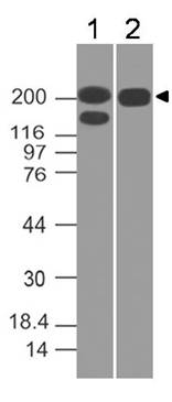
Figure-1: Western blot analysis of Spectrin beta 3. Anti-Spectrin beta 3 antibody (Clone: ABM5A16) was used at 0.1 µg/ml on h Kidney and h Brain lysates.
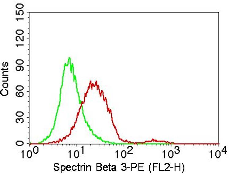
Figure-2: Intracellular flow analysis of human Spectrin beta 3 in A431 cells using 2 µg/10 6 cells of antibody (Clone: ABM5A16). Green represents isotype control, red represents anti-Spectrin beta 3 antibody. Goat anti-mouse PE conjugate was used as secondary antibody. (Cells were fixed with 4% paraformaldehyde for 10 min and washed with PBS by centrifuging at 1100 for 5 min followed by permeabilization for 20 min and washed again as mentioned above. Then cell were incubated with primary antibo
Figure-3: Intracellular flow analysis of human Spectrin beta 3 in HeLa cells using 2 µg/10 6 cells of antibody (Clone: ABM5A16). Green represents isotype control, red represents anti-Spectrin beta 3 antibody. Goat anti-mouse PE conjugate was used as secondary antibody. (Cells were fixed with 4% paraformaldehyde for 10 min and washed with PBS by centrifuging at 1100 for 5 min followed by permeabilization for 20 min and washed again as mentioned above. Then cell were incubated with primary antibo
* Mehrwertsteuer und Versandkosten nicht enthalten. Irrtümer und Preisänderungen vorbehalten