Monoclonal Antibody to Dc-Sign/CD 209 (Clone: ABM47B7)
Artikelnummer:
ABI-10-10020
Hersteller Artikelnummer:
10-10020
Alternativnummer:
ABI-10-10020-100UG
Hersteller:
Abeomics
Wirt:
Mouse
Kategorie:
Antikörper
Applikation:
FACS, WB
Spezies Reaktivität:
Human
Immunogen:
A recombinant protein fragment of DC-Sign protein was used as the immunogen for this antibody.
Alternative Synonym:
CD209||CLEC4L
Dendritic cell-specific intercellular adhesion molecule-3-grabbing non-integrin (DC-SIGN) is a tetrameric C-type (calcium-dependent) lectin that binds, through its C-terminal carbohydrate recognition domain, high mannose N-linked glycans present on the surface of several viral glycoproteins such as human immunodeficiency virus (HIV) gp120 and hepatitis C virus (HCV) E2. It facilitates DC-specific delivery of Ag. This is accomplished by conjugating Ag to receptor-specific Ab or carbohydrate ligands that bind to its carbohydrate recognition domain. In humans, DC-SIGN expression is restricted to DCs and certain types of macrophages. DC-SIGN is involved in the innate immune system and recognizes numerous evolutionarily divergent pathogens, including viruses, bacteria, fungi, and parasites. After binding, these pathogens are internalized and pathogen-derived antigens are presented via MHC class I and II molecules to CD8+ and CD4+ T cells, respectively. DC-SIGN represents a promising CLR for targeted vaccine delivery.
Western blot analysis: 0.1-0.5 µg/ml, FACS: 0.5-1 µg/10 6 Cells Reference for expression of Dc-Sign: Wai K. Lai, Phoebe J. Sun, Jie Zhang, Adam Jennings, Patricia F. Lalor, Stefan Hubscher,Jane A. McKeating,and David H. Adams. Expression of DC-SIGN and DC
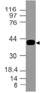
Fig-1: Expression analysis of Dc-Sign. Anti-Dc-Sign antibody (Clone: ABM47B7) was tested at 0.1 µg/ml on partial length recombinant protein.
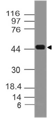
Fig-2: Expression analysis of Dc-Sign. Anti-Dc-Sign antibody (Clone: ABM47B7) was tested at 0.1 µg/ml on human Liver lysate.
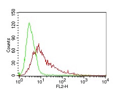
Fig-3: Cell Surface FLOW analysis of Dc-Sign antibody (10-10020) in 293HEK-Dc-sign stable cell line using 0.5 µg/ 10 6 cells. Green represents isotype control, red represents anti- Dc-Sign antibody. Goat anti-mouse PE conjugates was used as secondary antibody.
* Mehrwertsteuer und Versandkosten nicht enthalten. Irrtümer und Preisänderungen vorbehalten