
beta-Tubulin Rabbit pAb, Unconjugated
Artikelnummer:
ABB-AC015
- Bilder (9)


| Artikelname: | beta-Tubulin Rabbit pAb, Unconjugated |
| Artikelnummer: | ABB-AC015 |
| Hersteller Artikelnummer: | AC015 |
| Alternativnummer: | ABB-AC015-50UL,ABB-AC015-100UL,ABB-AC015-200UL,ABB-AC015-1000UL,ABB-AC015-500UL |
| Hersteller: | ABclonal |
| Wirt: | Rabbit |
| Kategorie: | Antikörper |
| Applikation: | IF, IHC-P, WB |
| Spezies Reaktivität: | Human |
| Immunogen: | Recombinant protein (or fragment).This information is considered to be commercially sensitive. |
| Konjugation: | Unconjugated |
| Alternative Synonym: | M40, TUBB1, TUBB5, CDCBM6, CSCSC1, OK/SW-cl.56, beta-Tubulin |
| This gene encodes a beta tubulin protein. This protein forms a dimer with alpha tubulin and acts as a structural component of microtubules. Mutations in this gene cause cortical dysplasia, complex, with other brain malformations 6. Alternative splicing results in multiple splice variants. There are multiple pseudogenes for this gene on chromosomes 1, 6, 7, 8, 9, and 13. |
| Klonalität: | Polyclonal |
| Molekulargewicht: | 50kDa |
| NCBI: | 203068 |
| UniProt: | P07437 |
| Reinheit: | Affinity purification |
| Sequenz: | SDLQLERISVYYNEASSHKYVPRAILVDLEPGTMDSVRSGAFGHLFRPDNFIFGQSGAGNNWAKGHYTEGAELVDSVLDVVRKECENCDCLQGFQLTHSLGGGTGSGMGTLLISKVREEYPDRIMNTFSVVPSPKVSDTVVEPYNATLSIHQLVENTDETYCIDNEALYDICFRTLKLATPTYGDLNHLVSATMSGVTTSLRFPGQLNADLRKLAVNMVPF |
| Target-Kategorie: | TUBB |
| Antibody Type: | Primary Antibody |
| Application Verdünnung: | WB,1:500 - 1:5000|IF/ICC,1:50 - 1:200|IHC-P,1:50 - 1:200 |
| Anwendungsbeschreibung: | Cross-Reactivity: Human,Mouse,Rat. ResearchArea: Cell Cycle,Centrosome,Cytoskeleton,Microtubules. Shipping: Ice Bag |
|
|
Western blot analysis of various lysates, using beta-Tubulin Rabbit pAb (AC015) at 1:2000 dilution. Secondary antibody: HRP-conjugated Goat anti-Rabbit IgG (H+L) (AS014) at 1:10000 dilution. Lysates/proteins: 25µg per lane. Blocking buffer: 3% nonfat dry milk in TBST. Detection: ECL Basic Kit (RM00020). Exposure time: 1s. |
|
|
Immunohistochemistry analysis of paraffin-embeddedRat brain tissue usingbeta-Tubulin Rabbit pAb(AC015) at a dilution of 1:100 (40x lens).High pressure antigen retrieval was performed with 0.01 M citrate buffer (pH 6.0) prior to IHC staining. |
|
|
Western blot analysis of various lysates using beta-Tubulin Rabbit pAb (AC015) at 1:1000 dilution. Secondary antibody: HRP-conjugated Goat anti-Rabbit IgG (H+L) (AS014) at 1:10000 dilution. Lysates/proteins: 25µg per lane. Blocking buffer: 3% nonfat dry milk in TBST. Detection: ECL Basic Kit (RM00020). Exposure time: 3s. |
|
|
Immunohistochemistry analysis of paraffin-embeddedMouse testis tissue usingbeta-Tubulin Rabbit pAb(AC015) at a dilution of 1:100 (40x lens).High pressure antigen retrieval was performed with 0.01 M citrate buffer (pH 6.0) prior to IHC staining. |
|
|
Immunohistochemistry analysis of paraffin-embeddedHuman colon tissue usingbeta-Tubulin Rabbit pAb(AC015) at a dilution of 1:100 (40x lens).High pressure antigen retrieval was performed with 0.01 M citrate buffer (pH 6.0) prior to IHC staining. |
|
|
Immunohistochemistry analysis of paraffin-embeddedHuman liver tissue usingbeta-Tubulin Rabbit pAb(AC015) at a dilution of 1:100 (40x lens).High pressure antigen retrieval was performed with 0.01 M citrate buffer (pH 6.0) prior to IHC staining. |
|
|
Immunohistochemistry analysis of paraffin-embeddedHuman cervix cancer tissue usingbeta-Tubulin Rabbit pAb(AC015) at a dilution of 1:100 (40x lens).High pressure antigen retrieval was performed with 0.01 M citrate buffer (pH 6.0) prior to IHC staining. |
|
|
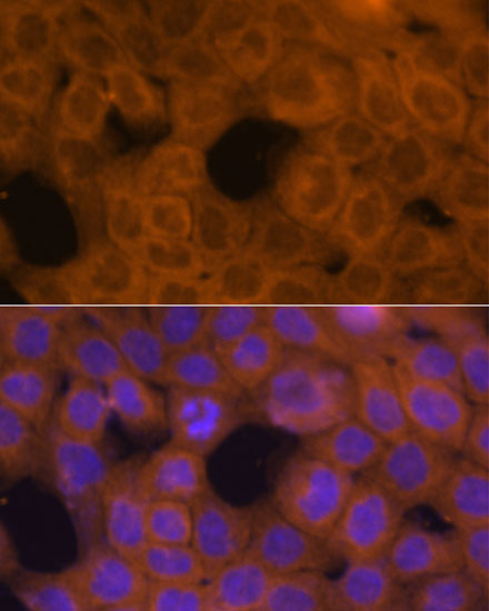
Immunofluorescence analysis of A431 cells using beta-Tubulin Rabbit pAb (AC015) at dilution of 1:100. Secondary antibody: Cy3-conjugated Goat anti-Rabbit IgG (H+L) (AS007) at 1:500 dilution. Blue: DAPI for nuclear staining. |
|
|
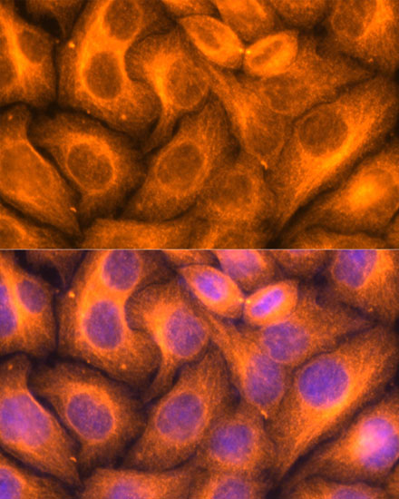
Immunofluorescence analysis of HeLa cells using beta-Tubulin Rabbit pAb (AC015) at dilution of 1:100. Secondary antibody: Cy3-conjugated Goat anti-Rabbit IgG (H+L) (AS007) at 1:500 dilution. Blue: DAPI for nuclear staining. |
Produktgarantie und fachkundiger Support
