
RPS20 Rabbit mAb, Unconjugated, Monoclonal
Artikelnummer:
ABB-A9551
- Bilder (8)

| Artikelname: | RPS20 Rabbit mAb, Unconjugated, Monoclonal |
| Artikelnummer: | ABB-A9551 |
| Hersteller Artikelnummer: | A9551 |
| Alternativnummer: | ABB-A9551-20UL,ABB-A9551-100UL |
| Hersteller: | ABclonal |
| Wirt: | Rabbit |
| Kategorie: | Antikörper |
| Applikation: | ELISA, IF, IHC-P, IP, WB |
| Spezies Reaktivität: | Human |
| Immunogen: | Synthetic peptide. This information is considered to be commercially sensitive. |
| Konjugation: | Unconjugated |
| Alternative Synonym: | S20, uS10, RPS20 |
| Ribosomes, the organelles that catalyze protein synthesis, consist of a small 40S subunit and a large 60S subunit. Together these subunits are composed of 4 RNA species and approximately 80 structurally distinct proteins. This gene encodes a ribosomal protein that is a component of the 40S subunit. The protein belongs to the S10P family of ribosomal proteins. It is located in the cytoplasm. This gene is co-transcribed with the small nucleolar RNA gene U54, which is located in its second intron. As is typical for genes encoding ribosomal proteins, there are multiple processed pseudogenes of this gene dispersed through the genome. Two transcript variants encoding different isoforms have been identified for this gene. |
| Application Verdünnung: | WB,1:500 - 1:1000|IHC-P,1:500 - 1:1000|IF/ICC,1:50 - 1:200|IP,0.5µg-4µg antibody for 200µg-400µg extracts of whole cells|ELISA,Recommended starting concentration is 1 µg/mL. Please optimize the concentration based on your specific assay requirements. |
| Anwendungsbeschreibung: | Cross-Reactivity: Human,Mouse,Rat. ResearchArea: Epigenetics Nuclear Signaling,RNA Binding. Shipping: Ice Bag |
|
|
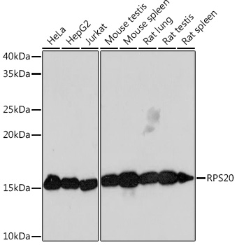
Western blot analysis of various lysates using RPS20 Rabbit mAb (A9551) at 1:1000 dilution. Secondary antibody: HRP-conjugated Goat anti-Rabbit IgG (H+L) (AS014) at 1:10000 dilution. Lysates/proteins: 25µg per lane. Blocking buffer: 3% nonfat dry milk in TBST. Detection: ECL Basic Kit (RM00020). Exposure time: 3min. |
|
|
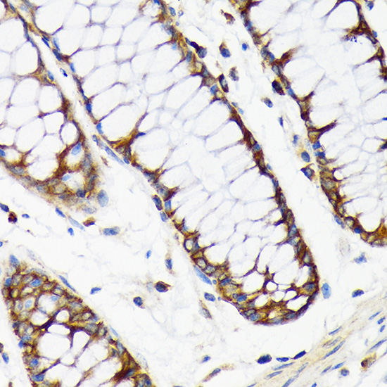
Immunohistochemistry analysis of paraffin-embedded Human colon carcinoma tissue using RPS20 Rabbit mAb (A9551) at a dilution of 1:800 (40x lens). High pressure antigen retrieval performed with 0.01M Citrate buffer (pH 6.0) prior to IHC staining. |
|
|
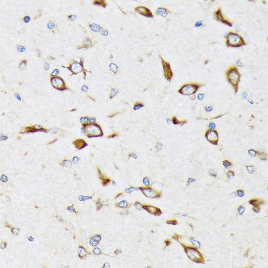
Immunohistochemistry analysis of paraffin-embedded Mouse brain tissue using RPS20 Rabbit mAb (A9551) at a dilution of 1:800 (40x lens). High pressure antigen retrieval performed with 0.01M Citrate buffer (pH 6.0) prior to IHC staining. |
|
|
Immunohistochemistry analysis of paraffin-embedded Mouse testis tissue using RPS20 Rabbit mAb (A9551) at a dilution of 1:800 (40x lens). High pressure antigen retrieval performed with 0.01M Citrate buffer (pH 6.0) prior to IHC staining. |
|
|
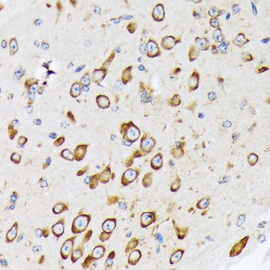
Immunohistochemistry analysis of paraffin-embedded Rat brain tissue using RPS20 Rabbit mAb (A9551) at a dilution of 1:800 (40x lens). High pressure antigen retrieval performed with 0.01M Citrate buffer (pH 6.0) prior to IHC staining. |
|
|
Immunohistochemistry analysis of paraffin-embedded Rat colon tissue using RPS20 Rabbit mAb (A9551) at a dilution of 1:800 (40x lens). High pressure antigen retrieval performed with 0.01M Citrate buffer (pH 6.0) prior to IHC staining. |
|
|
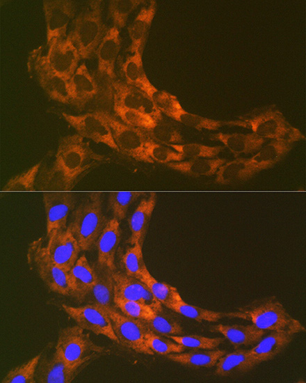
Immunofluorescence analysis of C6 cells using RPS20 Rabbit mAb (A9551) at dilution of 1:100 (40x lens). Secondary antibody: Cy3-conjugated Goat anti-Rabbit IgG (H+L) (AS007) at 1:500 dilution. Blue: DAPI for nuclear staining. |
|
|
Immunoprecipitation of RPS20 in 300 µg extracts from Hep G2 cells using 3 µg RPS20 Rabbit mAb (A9551). Western blot analysis was performed using RPS20 Rabbit mAb (A9551) at 1:1000 dilution. |
Produktgarantie und fachkundiger Support
