
EIF3F Rabbit pAb, Unconjugated, Polyclonal
Artikelnummer:
ABB-A7023
- Bilder (8)


| Artikelname: | EIF3F Rabbit pAb, Unconjugated, Polyclonal |
| Artikelnummer: | ABB-A7023 |
| Hersteller Artikelnummer: | A7023 |
| Alternativnummer: | ABB-A7023-20UL,ABB-A7023-100UL,ABB-A7023-500UL,ABB-A7023-1000UL |
| Hersteller: | ABclonal |
| Wirt: | Rabbit |
| Kategorie: | Antikörper |
| Applikation: | ELISA, IF, IHC-P, IP, WB |
| Spezies Reaktivität: | Human |
| Immunogen: | Recombinant protein (or fragment).This information is considered to be commercially sensitive. |
| Konjugation: | Unconjugated |
| Alternative Synonym: | MRT67, EIF3S5, eIF3-p47, EIF3F |
| Enables deubiquitinase activity and identical protein binding activity. Contributes to translation initiation factor activity. Involved in IRES-dependent viral translational initiation, protein deubiquitination, and translational initiation. Located in membrane. Part of eukaryotic translation initiation factor 3 complex. Implicated in autosomal recessive non-syndromic intellectual disability. |
| Klonalität: | Polyclonal |
| Molekulargewicht: | 38kDa |
| NCBI: | 8665 |
| UniProt: | O00303 |
| Reinheit: | Affinity purification |
| Sequenz: | GGRVVRLHPVILASIVDSYERRNEGAARVIGTLLGTVDKHSVEVTNCFSVPHNESEDEVAVDMEFAKNMYELHKKVSPNELILGWYATGHDITEHSVLIHEYYSREAPNPIHLTVDTSLQNGRMSIKAYVSTLMGVPGRTMGVMFTPLTVKYAYYDTERIGVDLIMKTCFSPNRVIGLSSDLQQVGGASARIQDALSTVLQYAEDVLSGKVSADNTVGRFLMSLVNQVPKIVPDDFETMLNSNINDLLMVTYLAN |
| Target-Kategorie: | EIF3F |
| Antibody Type: | Primary Antibody |
| Application Verdünnung: | WB,1:500 - 1:1000|IHC-P,1:50 - 1:200|IF/ICC,1:50 - 1:200|IP,0.5µg-4µg antibody for 200µg-400µg extracts of whole cells|ELISA,Recommended starting concentration is 1 µg/mL. Please optimize the concentration based on your specific assay requirements. |
| Anwendungsbeschreibung: | Cross-Reactivity: Human,Mouse,Rat. ResearchArea: Epigenetics Nuclear Signaling. Shipping: Ice Bag |
|
|
Immunohistochemistry analysis of paraffin-embeddedHuman colon tissue usingEIF3F Rabbit pAb(A7023) at a dilution of1:200 (40x lens).High pressure antigen retrieval was performed with 0.01 M citrate buffer (pH 6.0) prior to IHC staining. |
|
|
Immunohistochemistry analysis of paraffin-embeddedHuman pancreas tissue usingEIF3F Rabbit pAb(A7023) at a dilution of1:200 (40x lens).High pressure antigen retrieval was performed with 0.01 M citrate buffer (pH 6.0) prior to IHC staining. |
|
|
Western blot analysis of lysates from HeLa cells, using EIF3F Rabbit pAb (A7023) at 1:1000 dilution. Secondary antibody: HRP-conjugated Goat anti-Rabbit IgG (H+L) (AS014) at 1:10000 dilution. Lysates/proteins: 25µg per lane. Blocking buffer: 3% nonfat dry milk in TBST. Detection: ECL Enhanced Kit (RM00021). Exposure time: 60s. |
|
|
Immunohistochemistry analysis of paraffin-embeddedRat brain tissue usingEIF3F Rabbit pAb(A7023) at a dilution of1:200 (40x lens).High pressure antigen retrieval was performed with 0.01 M citrate buffer (pH 6.0) prior to IHC staining. |
|
|
Immunohistochemistry analysis of paraffin-embeddedMouse spleen tissue usingEIF3F Rabbit pAb(A7023) at a dilution of1:200 (40x lens).High pressure antigen retrieval was performed with 0.01 M citrate buffer (pH 6.0) prior to IHC staining. |
|
|
Immunofluorescence analysis of NIH/3T3 cells using EIF3F Rabbit pAb (A7023) at dilution of 1:200 (40x lens). Secondary antibody: Cy3-conjugated Goat anti-Rabbit IgG (H+L) (AS007) at 1:500 dilution. Blue: DAPI for nuclear staining. |
|
|
Immunofluorescence analysis of PC-12 cells using EIF3F Rabbit pAb (A7023) at dilution of 1:200 (40x lens). Secondary antibody: Cy3-conjugated Goat anti-Rabbit IgG (H+L) (AS007) at 1:500 dilution. Blue: DAPI for nuclear staining. |
|
|
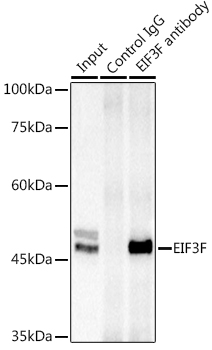
Immunoprecipitation analysis of 300 µg extracts of Jurkat cells using 3 µg EIF3F antibody (A7023). Western blot was performed from the immunoprecipitate using EIF3F antibody (A7023) at a dilution of 1:1000. |
Produktgarantie und fachkundiger Support
