
HTATSF1 Rabbit pAb, Unconjugated, Polyclonal
Artikelnummer:
ABB-A5977
- Bilder (9)


| Artikelname: | HTATSF1 Rabbit pAb, Unconjugated, Polyclonal |
| Artikelnummer: | ABB-A5977 |
| Hersteller Artikelnummer: | A5977 |
| Alternativnummer: | ABB-A5977-100UL,ABB-A5977-20UL,ABB-A5977-500UL,ABB-A5977-1000UL |
| Hersteller: | ABclonal |
| Wirt: | Rabbit |
| Kategorie: | Antikörper |
| Applikation: | ELISA, IF, IHC-P, WB |
| Spezies Reaktivität: | Human |
| Immunogen: | Recombinant protein (or fragment).This information is considered to be commercially sensitive. |
| Konjugation: | Unconjugated |
| Alternative Synonym: | TATSF1, TAT-SF1, dJ196E23.2, HTATSF1 |
| The protein encoded by this gene functions as a cofactor for the stimulation of transcriptional elongation by HIV-1 Tat, which binds to the HIV-1 promoter through Tat-TAR interaction. This protein may also serve as a dual-function factor to couple transcription and splicing and to facilitate their reciprocal activation. Alternatively spliced transcript variants have been found for this gene. |
| Klonalität: | Polyclonal |
| Molekulargewicht: | 86kDa |
| NCBI: | 27336 |
| UniProt: | O43719 |
| Reinheit: | Affinity purification |
| Sequenz: | MSGTNLDGNDEFDEQLRMQELYGDGKDGDTQTDAGGEPDSLGQQPTDTPYEWDLDKKAWFPKITEDFIATYQANYGFSNDGASSSTANVEDVHARTAEEPPQEKAPEPTDARKKGEKRKAESGWFHVEEDRNTNVYVSGLPPDITVDEFIQLMSKFGIIMRDPQTEEFKVKLYKDNQGNLKGDGLCCYLKRESVELALKLLDEDEIRGYKLHVEVAKFQLKGEYDASKKKKKCKDYKKKLSMQQKQLDWRPERRA |
| Target-Kategorie: | HTATSF1 |
| Antibody Type: | Primary Antibody |
| Application Verdünnung: | WB,1:1000 - 1:2000|IHC-P,1:50 - 1:100|IF/ICC,1:50 - 1:200|ELISA,Recommended starting concentration is 1 µg/mL. Please optimize the concentration based on your specific assay requirements. |
| Anwendungsbeschreibung: | Cross-Reactivity: Human,Mouse,Rat. ResearchArea: Epigenetics Nuclear Signaling,RNA Binding,Immunology Inflammation. Shipping: Ice Bag |
|
|
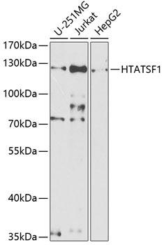
Western blot analysis of various lysates using HTATSF1 Rabbit pAb (A5977) at 1:1000 dilution. Secondary antibody: HRP-conjugated Goat anti-Rabbit IgG (H+L) (AS014) at 1:10000 dilution. Lysates/proteins: 25µg per lane. Blocking buffer: 3% nonfat dry milk in TBST. Detection: ECL Basic Kit (RM00020). Exposure time: 90s. |
|
|
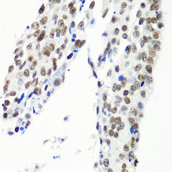
Immunohistochemistry analysis of paraffin-embedded Human lung cancer using HTATSF1 Rabbit pAb (A5977) at dilution of 1:100 (40x lens). Microwave antigen retrieval performed with 0.01M PBS Buffer (pH 7.2) prior to IHC staining. |
|
|
Western blot analysis of various lysates using HTATSF1 Rabbit pAb (A5977) at 1:1000 dilution. Secondary antibody: HRP-conjugated Goat anti-Rabbit IgG (H+L) (AS014) at 1:10000 dilution. Lysates/proteins: 25µg per lane. Blocking buffer: 3% nonfat dry milk in TBST. Detection: ECL Basic Kit (RM00020). Exposure time: 90s. |
|
|
Immunohistochemistry analysis of paraffin-embedded Human stomach using HTATSF1 Rabbit pAb (A5977) at dilution of 1:100 (40x lens). Microwave antigen retrieval performed with 0.01M PBS Buffer (pH 7.2) prior to IHC staining. |
|
|
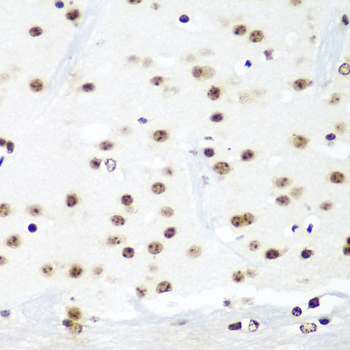
Immunohistochemistry analysis of paraffin-embedded Mouse brain using HTATSF1 Rabbit pAb (A5977) at dilution of 1:100 (40x lens). Microwave antigen retrieval performed with 0.01M PBS Buffer (pH 7.2) prior to IHC staining. |
|
|
Immunohistochemistry analysis of paraffin-embedded Human lung cancer using HTATSF1 Rabbit pAb (A5977) at dilution of 1:100 (40x lens). Microwave antigen retrieval performed with 0.01M PBS Buffer (pH 7.2) prior to IHC staining. |
|
|
Immunohistochemistry analysis of paraffin-embedded Human gastric cancer using HTATSF1 Rabbit pAb (A5977) at dilution of 1:100 (40x lens). Microwave antigen retrieval performed with 0.01M PBS Buffer (pH 7.2) prior to IHC staining. |
|
|
Immunohistochemistry analysis of paraffin-embedded Mouse testis using HTATSF1 Rabbit pAb (A5977) at dilution of 1:100 (40x lens). Microwave antigen retrieval performed with 0.01M PBS Buffer (pH 7.2) prior to IHC staining. |
|
|
Immunofluorescence analysis of C6 cells using HTATSF1 Rabbit pAb (A5977) at dilution of 1:100. Secondary antibody: Cy3-conjugated Goat anti-Rabbit IgG (H+L) (AS007) at 1:500 dilution. Blue: DAPI for nuclear staining. |
Produktgarantie und fachkundiger Support
