
ROCK2 Rabbit pAb, Unconjugated, Polyclonal
Artikelnummer:
ABB-A5698
- Bilder (9)
| Artikelname: | ROCK2 Rabbit pAb, Unconjugated, Polyclonal |
| Artikelnummer: | ABB-A5698 |
| Hersteller Artikelnummer: | A5698 |
| Alternativnummer: | ABB-A5698-20UL,ABB-A5698-100UL,ABB-A5698-500UL,ABB-A5698-1000UL |
| Hersteller: | ABclonal |
| Wirt: | Rabbit |
| Kategorie: | Antikörper |
| Applikation: | ELISA, IF, IHC-P, WB |
| Spezies Reaktivität: | Human |
| Immunogen: | Recombinant protein (or fragment).This information is considered to be commercially sensitive. |
| Konjugation: | Unconjugated |
| Alternative Synonym: | ROCK-II, ROCK2 |
| The protein encoded by this gene is a serine/threonine kinase that regulates cytokinesis, smooth muscle contraction, the formation of actin stress fibers and focal adhesions, and the activation of the c-fos serum response element. This protein, which is an isozyme of ROCK1 is a target for the small GTPase Rho. |
| Klonalität: | Polyclonal |
| Molekulargewicht: | 161kDa |
| NCBI: | 9475 |
| UniProt: | O75116 |
| Reinheit: | Affinity purification |
| Sequenz: | LDSKDSDIEQLRSQLQALHIGLDSSSIGSGPGDAEADDGFPESRLEGWLSLPVRNNTKKFGWVKKYVIVSSKKILFYDSEQDKEQSNPYMVLDIDKLFHVRPVTQTDVYRADAKEIPRIFQILYANEGESKKEQEFPVEPVGEKSNYICHKGHEFIPTLYHFPTNCEACMKPLWHMFKPPPALECRRCHIKCHKDHMDKKEEIIAPCKVYYDISTAKNLLLLANSTEEQQKWVSRLVKKIPKKPPAPDPFARSSP |
| Target-Kategorie: | ROCK2 |
| Antibody Type: | Primary Antibody |
| Application Verdünnung: | WB,1:500 - 1:1000|IHC-P,1:50 - 1:200|IF/ICC,1:50 - 1:200|ELISA,Recommended starting concentration is 1 µg/mL. Please optimize the concentration based on your specific assay requirements. |
| Anwendungsbeschreibung: | Cross-Reactivity: Human,Mouse,Rat. ResearchArea: Cancer,Signal Transduction,G protein signaling,Kinase,Serine threonine kinases,Cell Biology Developmental Biology,Apoptosis,Cell Cycle,Centrosome,Cell Adhesion,Cytoskeleton,Microtubules,Actins,TGF-b-Smad Signaling Pathway. Shipping: Ice Bag |
|
|
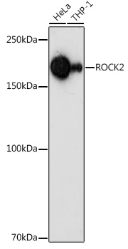
Western blot analysis of various lysates using ROCK2 Rabbit pAb (A5698) at 1:1000 dilution. Secondary antibody: HRP-conjugated Goat anti-Rabbit IgG (H+L) (AS014) at 1:10000 dilution. Lysates/proteins: 25µg per lane. Blocking buffer: 3% nonfat dry milk in TBST. Detection: ECL Basic Kit (RM00020). Exposure time: 180s. |
|
|
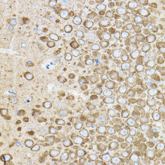
Immunohistochemistry analysis of paraffin-embedded Rat brain using ROCK2 Rabbit pAb (A5698) at dilution of 1:50 (40x lens). High pressure antigen retrieval performed with 0.01M Citrate buffer (pH 6.0) prior to IHC staining. |
|
|
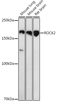
Western blot analysis of various lysates using ROCK2 Rabbit pAb (A5698) at 1:1000 dilution. Secondary antibody: HRP-conjugated Goat anti-Rabbit IgG (H+L) (AS014) at 1:10000 dilution. Lysates/proteins: 25µg per lane. Blocking buffer: 3% nonfat dry milk in TBST. Detection: ECL Basic Kit (RM00020). Exposure time: 180s. |
|
|
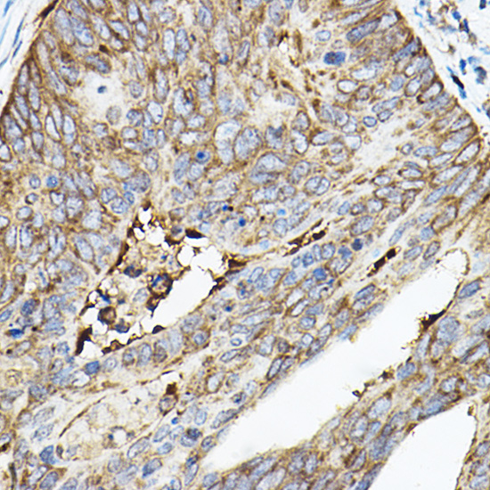
Immunohistochemistry analysis of paraffin-embedded Human colon carcinoma using ROCK2 Rabbit pAb (A5698) at dilution of 1:50 (40x lens). High pressure antigen retrieval performed with 0.01M Citrate buffer (pH 6.0) prior to IHC staining. |
|
|
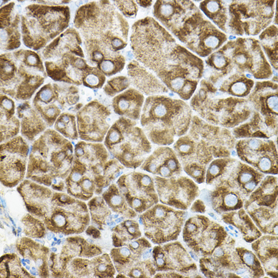
Immunohistochemistry analysis of paraffin-embedded Mouse pancreas using ROCK2 Rabbit pAb (A5698) at dilution of 1:50 (40x lens). High pressure antigen retrieval performed with 0.01M Citrate buffer (pH 6.0) prior to IHC staining. |
|
|
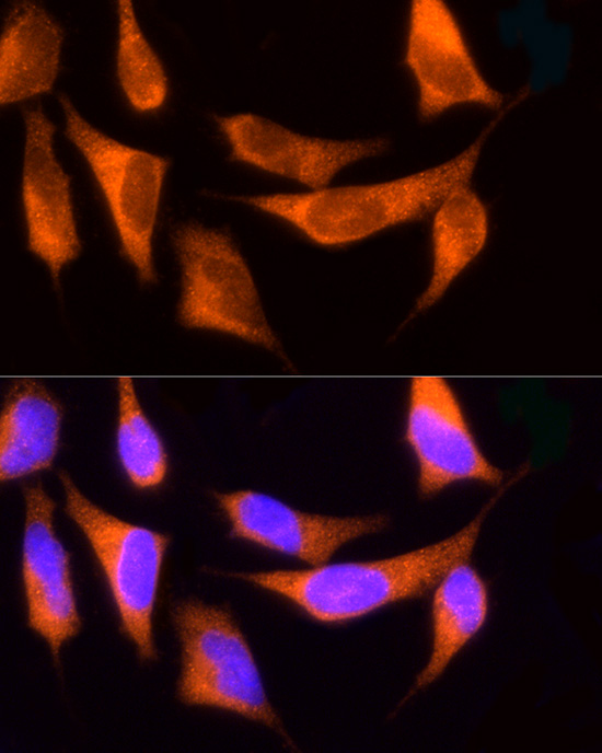
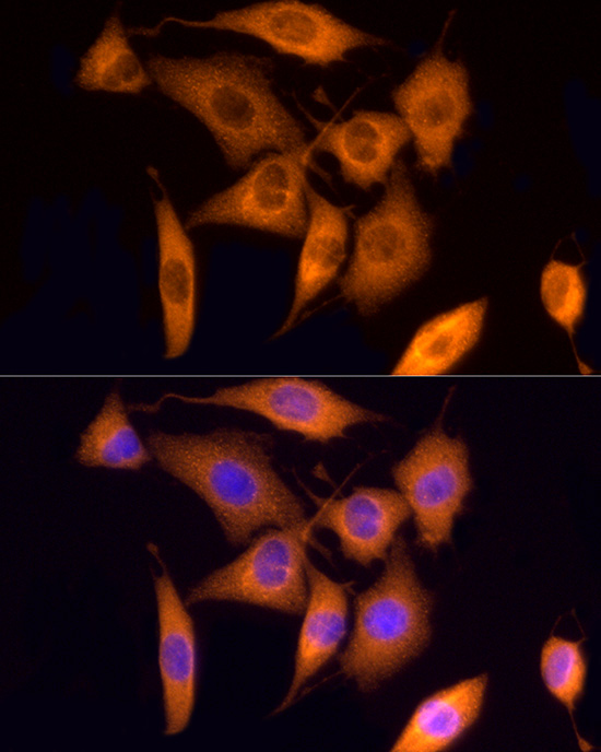
Immunofluorescence analysis of HeLa cells using ROCK2 Rabbit pAb (A5698) at dilution of 1:100 (40x lens). Secondary antibody: Cy3-conjugated Goat anti-Rabbit IgG (H+L) (AS007) at 1:500 dilution. Blue: DAPI for nuclear staining. |
|
|
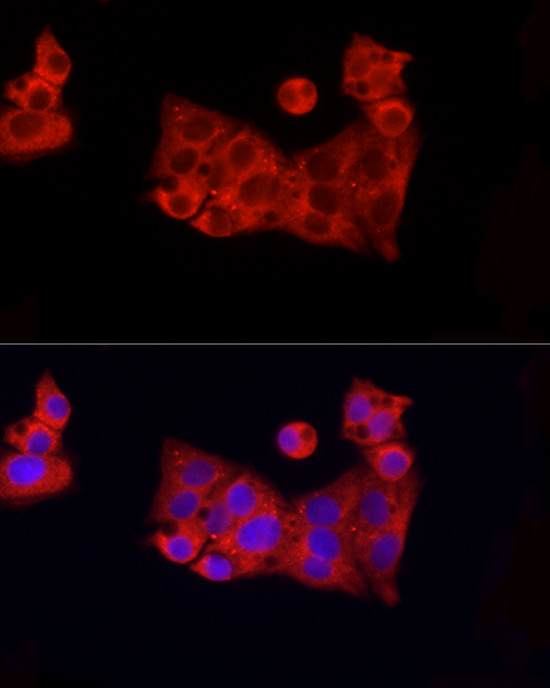
Immunofluorescence analysis of HepG2 cells using ROCK2 Rabbit pAb (A5698) at dilution of 1:100 (40x lens). Secondary antibody: Cy3-conjugated Goat anti-Rabbit IgG (H+L) (AS007) at 1:500 dilution. Blue: DAPI for nuclear staining. |
|
|
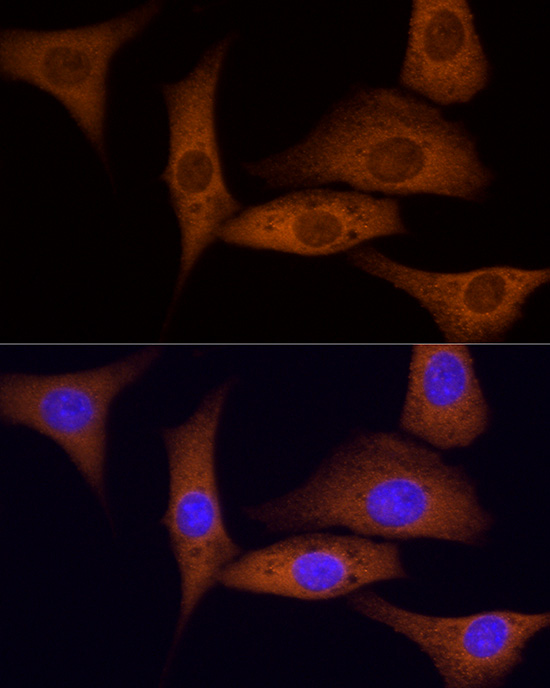
Immunofluorescence analysis of NIH/3T3 cells using ROCK2 Rabbit pAb (A5698) at dilution of 1:100 (40x lens). Secondary antibody: Cy3-conjugated Goat anti-Rabbit IgG (H+L) (AS007) at 1:500 dilution. Blue: DAPI for nuclear staining. |
|
|
Immunofluorescence analysis of PC-12 cells using ROCK2 Rabbit pAb (A5698) at dilution of 1:100 (40x lens). Secondary antibody: Cy3-conjugated Goat anti-Rabbit IgG (H+L) (AS007) at 1:500 dilution. Blue: DAPI for nuclear staining. |
Produktgarantie und fachkundiger Support
