
ERK1/2 Rabbit mAb, Unconjugated, Monoclonal
Artikelnummer:
ABB-A4782
- Bilder (9)


| Artikelname: | ERK1/2 Rabbit mAb, Unconjugated, Monoclonal |
| Artikelnummer: | ABB-A4782 |
| Hersteller Artikelnummer: | A4782 |
| Alternativnummer: | ABB-A4782-20UL,ABB-A4782-100UL,ABB-A4782-500UL,ABB-A4782-1000UL |
| Hersteller: | ABclonal |
| Wirt: | Rabbit |
| Kategorie: | Antikörper |
| Applikation: | ELISA, IF, IHC-P, WB |
| Spezies Reaktivität: | Human |
| Immunogen: | Recombinant protein (or fragment).This information is considered to be commercially sensitive. |
| Konjugation: | Unconjugated |
| Alternative Synonym: | ERK, ERK-2, ERK2, ERT1, MAPK2, P42MAPK, PRKM1, PRKM2, p38, p40, p41, p41mapk, p42-MAPK, 5594/5595, ERK1/2 |
| This gene encodes a member of the MAP kinase family. MAP kinases, also known as extracellular signal-regulated kinases (ERKs), act as an integration point for multiple biochemical signals, and are involved in a wide variety of cellular processes such as proliferation, differentiation, transcription regulation and development. The activation of this kinase requires its phosphorylation by upstream kinases. Upon activation, this kinase translocates to the nucleus of the stimulated cells, where it phosphorylates nuclear targets. One study also suggests that this protein acts as a transcriptional repressor independent of its kinase activity. The encoded protein has been identified as a moonlighting protein based on its ability to perform mechanistically distinct functions. Two alternatively spliced transcript variants encoding the same protein, but differing in the UTRs, have been reported for this gene. [provided by RefSeq, Jan 2014] |
| Klonalität: | Monoclonal |
| Klon-Bezeichnung: | [ARC0212] |
| Molekulargewicht: | 42 kDa/44 kDa |
| NCBI: | 5594 |
| UniProt: | P28482 |
| Reinheit: | Affinity purification |
| Sequenz: | HTGFLTEYVATRWYRAPEIMLNSKGYTKSIDIWSVGCILAEMLSNRPIFPGKHYLDQLNHILGILGSPSQEDLNCIINLKARNYLLSLPHKNKVPWNRLFPNADSKALDLLDKMLTFNPHKRIEVEQALAHPYLEQYYDPSDEPIAEAPFKFDMELDDLPKEKLKELIFEETARFQPGYRS |
| Target-Kategorie: | MAPK1/MAPK3 |
| Antibody Type: | Primary Antibody |
| Application Verdünnung: | WB,1:1000 - 1:6000|IHC-P,1:4000 - 1:16000|IF/ICC,1:200 - 1:400|ELISA,Recommended starting concentration is 1 µg/mL. Please optimize the concentration based on your specific assay requirements. |
| Anwendungsbeschreibung: | Cross-Reactivity: Human,Mouse,Rat. ResearchArea: Protein phosphorylation. Shipping: Ice Bag |
|
|
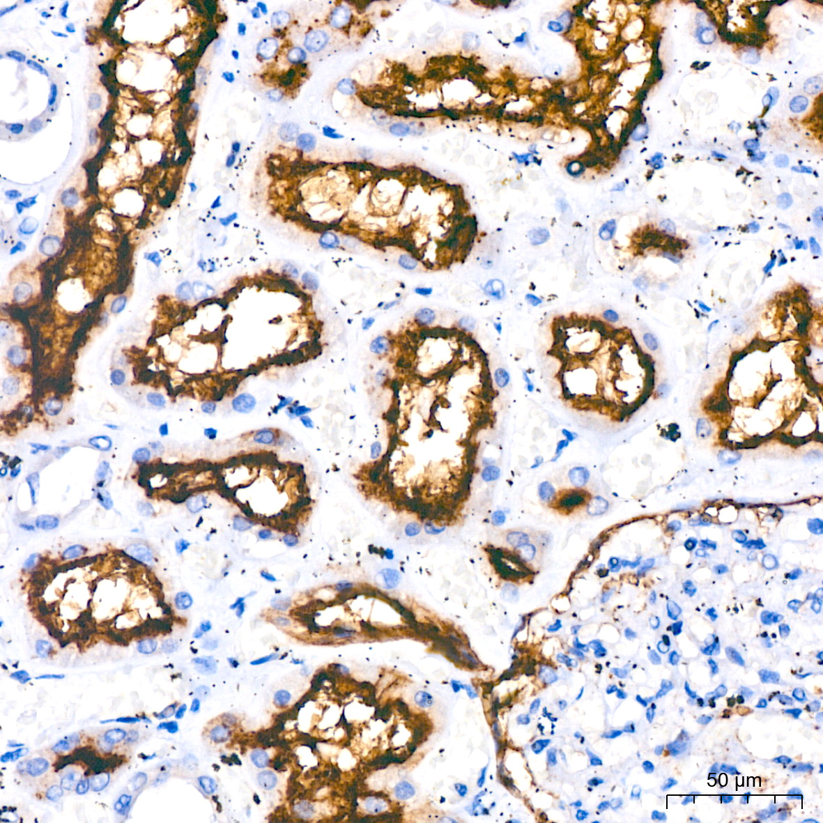
Immunohistochemistry analysis of paraffin-embedded Human kidney tissue using ERK1/2 Rabbit mAb (A4782) at a dilution of 1:9600 (40x lens). High pressure antigen retrieval performed with 0.01M Tris-EDTA Buffer (pH 9.0) prior to IHC staining. |
|
|
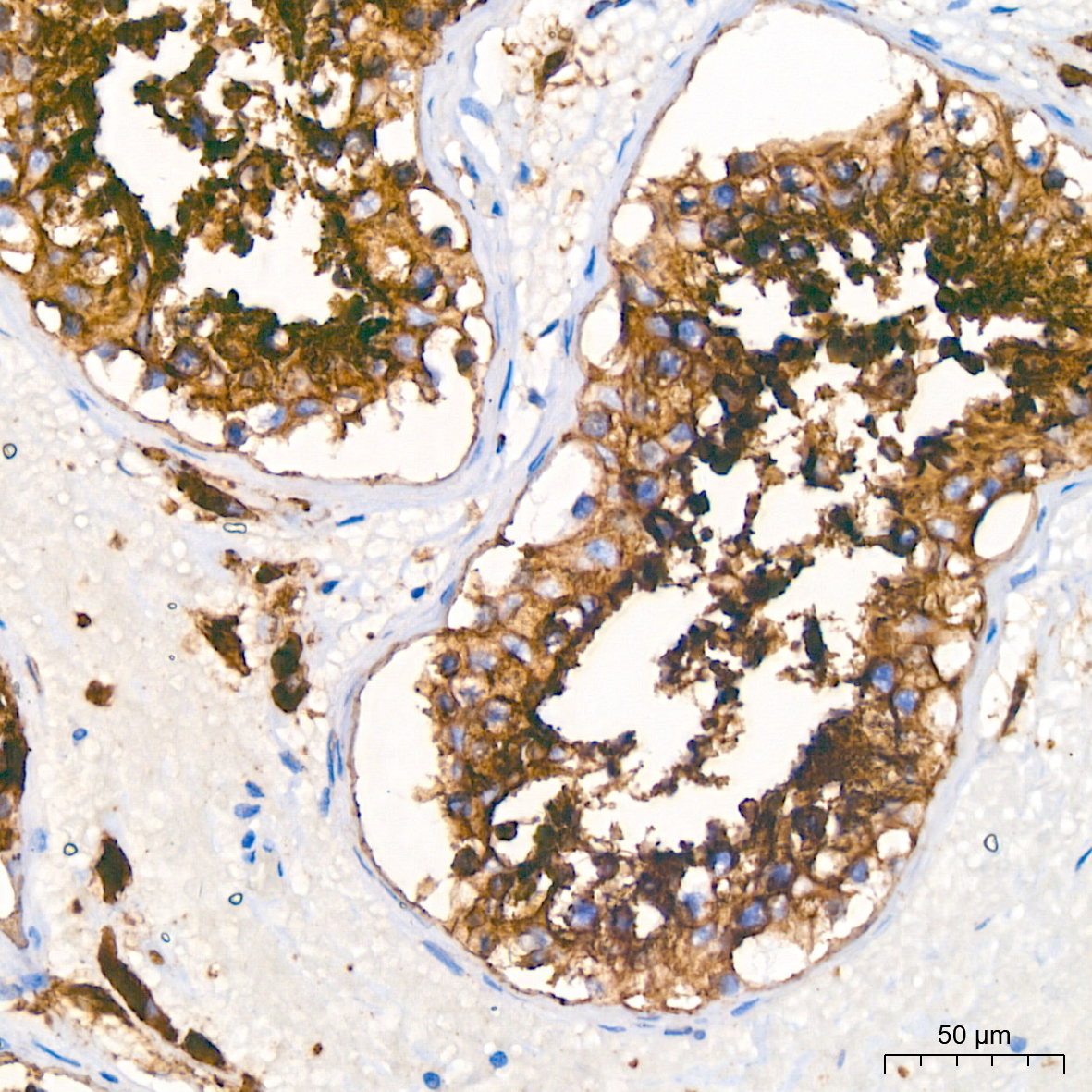
Immunohistochemistry analysis of paraffin-embedded Human colon carcinoma tissue using ERK1/2 Rabbit mAb (A4782) at a dilution of 1:9600 (40x lens). High pressure antigen retrieval performed with 0.01M Tris-EDTA Buffer (pH 9.0) prior to IHC staining. |
|
|
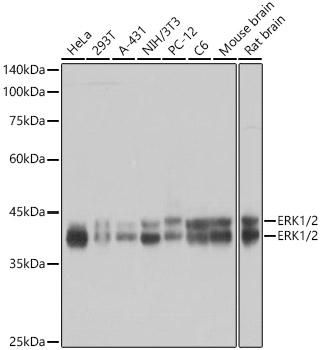
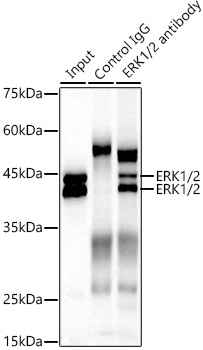
Western blot analysis of various lysates using ERK1/2 Rabbit mAb (A4782) at 1:4000 dilution incubated at room temperature for 1.5 hours. Secondary antibody: HRP-conjugated Goat anti-Rabbit IgG (H+L) (AS014) at 1:10000 dilution. Lysates/proteins: 25 µg per lane. Blocking buffer: 3% nonfat dry milk in TBST. Detection: ECL Basic Kit (RM00020). Exposure time: 45s. |
|
|
Immunohistochemistry analysis of paraffin-embedded Human tonsil tissue using ERK1/2 Rabbit mAb (A4782) at a dilution of 1:9600 (40x lens). High pressure antigen retrieval performed with 0.01M Tris-EDTA Buffer (pH 9.0) prior to IHC staining. |
|
|
Immunohistochemistry analysis of paraffin-embedded Mouse brain tissue using ERK1/2 Rabbit mAb (A4782) at a dilution of 1:9600 (40x lens). High pressure antigen retrieval performed with 0.01M Tris-EDTA Buffer (pH 9.0) prior to IHC staining. |
|
|
Immunohistochemistry analysis of paraffin-embedded Rat brain tissue using ERK1/2 Rabbit mAb (A4782) at a dilution of 1:9600 (40x lens). High pressure antigen retrieval performed with 0.01M Tris-EDTA Buffer (pH 9.0) prior to IHC staining. |
|
|
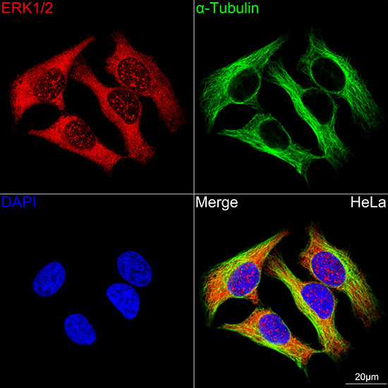
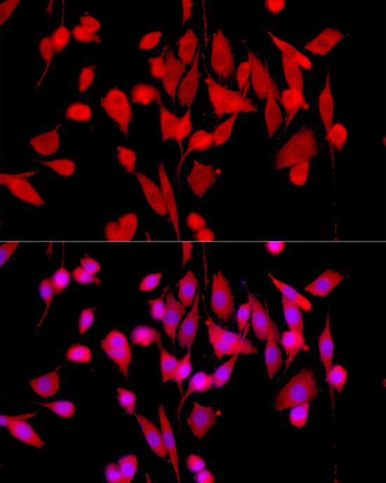
Confocal imaging of HeLa cells using ERK1/2 Rabbit mAb (A4782, dilution 1:200) followed by a further incubation with Cy3-conjugated Goat Anti-Rabbit IgG (H+L) (AS007, dilution 1:500) (Red). The cells were counterstained with alpha-Tubulin Mouse mAb (AC012, dilution 1:400) followed by incubation with ABflo 488-conjugated Goat Anti-Mouse IgG (H+L) (AS076, dilution 1:500) (Green). DAPI was used for nuclear staining (Blue). Objective: 100x. |
|
|
Confocal imaging of NIH/3T3 cells using ERK1/2 Rabbit mAb (A4782, dilution 1:200) followed by a further incubation with Cy3-conjugated Goat Anti-Rabbit IgG (H+L) (AS007, dilution 1:500) (Red). The cells were counterstained with alpha-Tubulin Mouse mAb (AC012, dilution 1:400) followed by incubation with ABflo 488-conjugated Goat Anti-Mouse IgG (H+L) (AS076, dilution 1:500) (Green). DAPI was used for nuclear staining (Blue). Objective: 100x. |
|
|
Confocal imaging of PC-12 cells using ERK1/2 Rabbit mAb (A4782, dilution 1:200) followed by a further incubation with Cy3-conjugated Goat Anti-Rabbit IgG (H+L) (AS007, dilution 1:500) (Red). The cells were counterstained with alpha-Tubulin Mouse mAb (AC012, dilution 1:400) followed by incubation with ABflo 488-conjugated Goat Anti-Mouse IgG (H+L) (AS076, dilution 1:500) (Green). DAPI was used for nuclear staining (Blue). Objective: 100x. |
Produktgarantie und fachkundiger Support
