
[KO Validated] KAT1/HAT1 Rabbit mAb, Unconjugated, Monoclonal
Artikelnummer:
ABB-A4423
- Bilder (9)

| Artikelname: | [KO Validated] KAT1/HAT1 Rabbit mAb, Unconjugated, Monoclonal |
| Artikelnummer: | ABB-A4423 |
| Hersteller Artikelnummer: | A4423 |
| Alternativnummer: | ABB-A4423-100UL,ABB-A4423-20UL |
| Hersteller: | ABclonal |
| Wirt: | Rabbit |
| Kategorie: | Antikörper |
| Applikation: | ELISA, IF, IHC-P, IP, WB |
| Spezies Reaktivität: | Human |
| Immunogen: | Recombinant protein (or fragment).This information is considered to be commercially sensitive. |
| Konjugation: | Unconjugated |
| Alternative Synonym: | KAT1, KAT1/HAT1 |
| The protein encoded by this gene is a type B histone acetyltransferase (HAT) that is involved in the rapid acetylation of newly synthesized cytoplasmic histones, which are in turn imported into the nucleus for de novo deposition onto nascent DNA chains. Histone acetylation, particularly of histone H4, plays an important role in replication-dependent chromatin assembly. Specifically, this HAT can acetylate soluble but not nucleosomal histone H4 at lysines 5 and 12, and to a lesser degree, histone H2A at lysine 5. Alternatively spliced transcript variants have been identified for this gene. |
| Application Verdünnung: | WB,1:1000 - 1:4000|IP,0.5µg-4µg antibody for 200µg-400µg extracts of whole cells|IHC-P,1:50 - 1:200|IF/ICC,1:50 - 1:200|ELISA,Recommended starting concentration is 1 µg/mL. Please optimize the concentration based on your specific assay requirements. |
| Anwendungsbeschreibung: | Cross-Reactivity: Human,Mouse,Rat. ResearchArea: Epigenetics Nuclear Signaling,Epigenetic writers and erasers of core Histones,Cell Biology Developmental Biology. Shipping: Ice Bag |
|
|
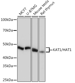
Western blot analysis of various lysates using [KO Validated] KAT1/HAT1 Rabbit mAb (A4423) at 1:1000 dilution. Secondary antibody: HRP-conjugated Goat anti-Rabbit IgG (H+L) (AS014) at 1:10000 dilution. Lysates/proteins: 25 µg per lane. Blocking buffer: 3% nonfat dry milk in TBST. Detection: ECL Basic Kit (RM00020). Exposure time: 10 s. |
|
|
Western blot analysis of lysates from wild type (WT) and KAT1/HAT1 knockout (KO) HeLa cells using KAT1/HAT1 Rabbit mAb (A4423) at 1:1000 dilution incubated at room temperature for 1.5 hours. Secondary antibody: HRP-conjugated Goat anti-Rabbit IgG (H+L) (AS014) at 1:10000 dilution. Lysates/proteins: 25 µg per lane. Blocking buffer: 3% nonfat dry milk in TBST. Detection: ECL Basic Kit (RM00020). Exposure time: 1 s. |
|
|
Immunohistochemistry analysis of paraffin-embedded Human cervix cancer tissue using [KO Validated] KAT1/HAT1 Rabbit mAb (A4423) at a dilution of 1:200 (40x lens). High pressure antigen retrieval performed with 0.01M Citrate buffer (pH 6.0) prior to IHC staining. |
|
|
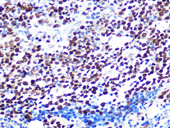
Immunohistochemistry analysis of paraffin-embedded Human colon tissue using [KO Validated] KAT1/HAT1 Rabbit mAb (A4423) at a dilution of 1:200 (40x lens). High pressure antigen retrieval performed with 0.01M Citrate buffer (pH 6.0) prior to IHC staining. |
|
|
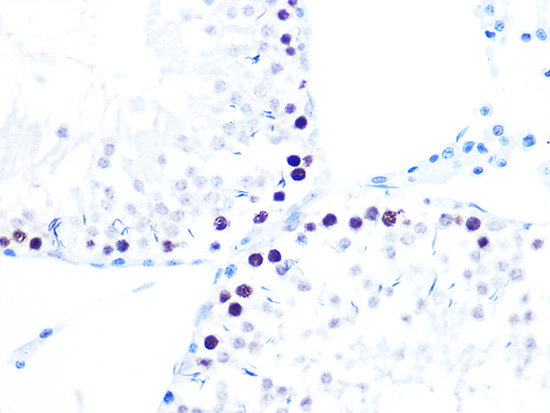
Immunohistochemistry analysis of paraffin-embedded Mouse testis tissue using [KO Validated] KAT1/HAT1 Rabbit mAb (A4423) at a dilution of 1:200 (40x lens). High pressure antigen retrieval performed with 0.01M Citrate buffer (pH 6.0) prior to IHC staining. |
|
|
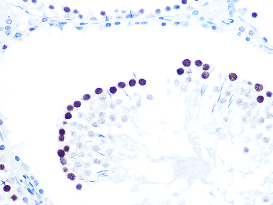
Immunohistochemistry analysis of paraffin-embedded Rat colon tissue using [KO Validated] KAT1/HAT1 Rabbit mAb (A4423) at a dilution of 1:200 (40x lens). High pressure antigen retrieval performed with 0.01M Citrate buffer (pH 6.0) prior to IHC staining. |
|
|
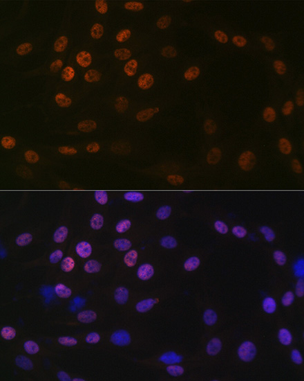
Immunofluorescence analysis of C6 cells using [KO Validated] KAT1/HAT1 Rabbit mAb (A4423) at dilution of 1:100 (40x lens). Secondary antibody: Cy3-conjugated Goat anti-Rabbit IgG (H+L) (AS007) at 1:500 dilution. Blue: DAPI for nuclear staining. |
|
|
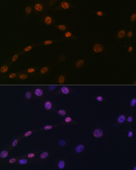
Immunofluorescence analysis of NIH/3T3 cells using [KO Validated] KAT1/HAT1 Rabbit mAb (A4423) at dilution of 1:100 (40x lens). Secondary antibody: Cy3-conjugated Goat anti-Rabbit IgG (H+L) (AS007) at 1:500 dilution. Blue: DAPI for nuclear staining. |
|
|
Immunoprecipitation of KAT1/HAT1 from 300 µg extracts of MCF7 cells was performed using 2 µg of [KO Validated] KAT1/HAT1 Rabbit mAb (A4423). Rabbit Control IgG (AC005) was used to precipitate the Control IgG sample. IP samples were eluted with 1X Laemmli Buffer. The Input lane represents 10% of the total input. Western blot analysis of immunoprecipitates was conducted using [KO Validated] KAT1/HAT1 Rabbit mAb (A4423) at a dilution of 1:1500. |
Produktgarantie und fachkundiger Support
