
DKC1 Rabbit mAb, Unconjugated, Monoclonal
Artikelnummer:
ABB-A4407
- Bilder (9)

| Artikelname: | DKC1 Rabbit mAb, Unconjugated, Monoclonal |
| Artikelnummer: | ABB-A4407 |
| Hersteller Artikelnummer: | A4407 |
| Alternativnummer: | ABB-A4407-100UL,ABB-A4407-20UL |
| Hersteller: | ABclonal |
| Wirt: | Rabbit |
| Kategorie: | Antikörper |
| Applikation: | ELISA, IHC-P, WB |
| Spezies Reaktivität: | Human |
| Immunogen: | Synthetic peptide. This information is considered to be commercially sensitive. |
| Konjugation: | Unconjugated |
| Alternative Synonym: | DKC, CBF5, DKCX, NAP57, NOLA4, XAP101, DKC1 |
| This gene functions in two distinct complexes. It plays an active role in telomerase stabilization and maintenance, as well as recognition of snoRNAs containing H/ACA sequences which provides stability during biogenesis and assembly into H/ACA small nucleolar RNA ribonucleoproteins (snoRNPs). This gene is highly conserved and widely expressed, and may play additional roles in nucleo-cytoplasmic shuttling, DNA damage response, and cell adhesion. Mutations have been associated with X-linked dyskeratosis congenita. Alternative splicing results in multiple transcript variants. |
| Application Verdünnung: | WB,1:500 - 1:2000|IHC-P,1:50 - 1:200|ELISA,Recommended starting concentration is 1 µg/mL. Please optimize the concentration based on your specific assay requirements. |
| Anwendungsbeschreibung: | Cross-Reactivity: Human,Mouse,Rat. ResearchArea: Epigenetics Nuclear Signaling,RNA Binding. Shipping: Ice Bag |
|
|
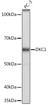
Western blot analysis of lysates from PC-3 cells, using DKC1 Rabbit mAb (A4407) at 1:1000 dilution. Secondary antibody: HRP-conjugated Goat anti-Rabbit IgG (H+L) (AS014) at 1:10000 dilution. Lysates/proteins: 25µg per lane. Blocking buffer: 3% nonfat dry milk in TBST. Detection: ECL Basic Kit (RM00020). Exposure time: 1s. |
|
|
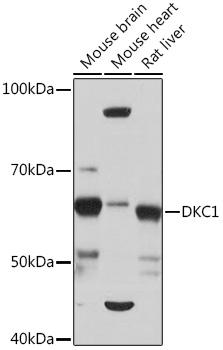
Western blot analysis of various lysates using DKC1 Rabbit mAb (A4407) at 1:1000 dilution. Secondary antibody: HRP-conjugated Goat anti-Rabbit IgG (H+L) (AS014) at 1:10000 dilution. Lysates/proteins: 25µg per lane. Blocking buffer: 3% nonfat dry milk in TBST. Detection: ECL Basic Kit (RM00020). Exposure time: 10s. |
|
|
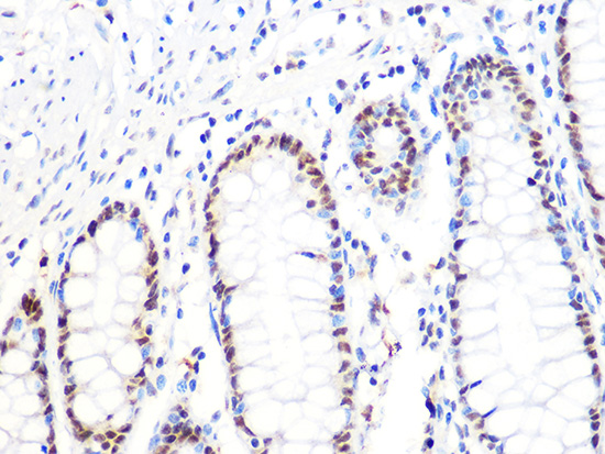
Immunohistochemistry analysis of paraffin-embeddedMouse colon tissue usingDKC1 Rabbit mAb(A4407) at a dilution of 1:200 (40x lens).High pressure antigen retrieval was performed with 0.01 M citrate buffer (pH 6.0) prior to IHC staining. |
|
|
Immunohistochemistry analysis of paraffin-embeddedHuman cervix cancer tissue usingDKC1 Rabbit mAb(A4407) at a dilution of 1:200 (40x lens).High pressure antigen retrieval was performed with 0.01 M citrate buffer (pH 6.0) prior to IHC staining. |
|
|
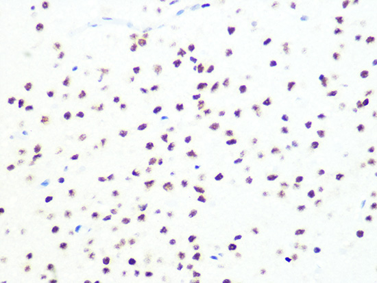
Immunohistochemistry analysis of paraffin-embeddedMouse brain tissue usingDKC1 Rabbit mAb(A4407) at a dilution of 1:200 (40x lens).High pressure antigen retrieval was performed with 0.01 M citrate buffer (pH 6.0) prior to IHC staining. |
|
|
Immunohistochemistry analysis of paraffin-embeddedRat colon tissue usingDKC1 Rabbit mAb(A4407) at a dilution of 1:200 (40x lens).High pressure antigen retrieval was performed with 0.01 M citrate buffer (pH 6.0) prior to IHC staining. |
|
|
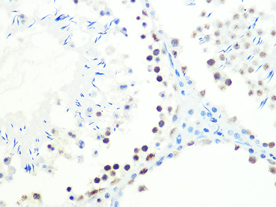
Immunohistochemistry analysis of paraffin-embeddedMouse testis tissue usingDKC1 Rabbit mAb(A4407) at a dilution of 1:200 (40x lens).High pressure antigen retrieval was performed with 0.01 M citrate buffer (pH 6.0) prior to IHC staining. |
|
|
Immunohistochemistry analysis of paraffin-embeddedRat spleen tissue usingDKC1 Rabbit mAb(A4407) at a dilution of 1:200 (40x lens).High pressure antigen retrieval was performed with 0.01 M citrate buffer (pH 6.0) prior to IHC staining. |
|
|
Immunohistochemistry analysis of paraffin-embeddedHuman spleen tissue usingDKC1 Rabbit mAb(A4407) at a dilution of 1:200 (40x lens).High pressure antigen retrieval was performed with 0.01 M citrate buffer (pH 6.0) prior to IHC staining. |
Produktgarantie und fachkundiger Support
