
VAMP2 Rabbit mAb, Unconjugated, Monoclonal
Artikelnummer:
ABB-A4235
- Bilder (9)

| Artikelname: | VAMP2 Rabbit mAb, Unconjugated, Monoclonal |
| Artikelnummer: | ABB-A4235 |
| Hersteller Artikelnummer: | A4235 |
| Alternativnummer: | ABB-A4235-20UL,ABB-A4235-100UL |
| Hersteller: | ABclonal |
| Wirt: | Rabbit |
| Kategorie: | Antikörper |
| Applikation: | ELISA, IF, IHC-P, WB |
| Spezies Reaktivität: | Human |
| Immunogen: | Synthetic peptide. This information is considered to be commercially sensitive. |
| Konjugation: | Unconjugated |
| Alternative Synonym: | SYB2, VAMP-2, NEDHAHM, VAMP2 |
| The protein encoded by this gene is a member of the vesicle-associated membrane protein (VAMP)/synaptobrevin family. Synaptobrevins/VAMPs, syntaxins, and the 25-kD synaptosomal-associated protein SNAP25 are the main components of a protein complex involved in the docking and/or fusion of synaptic vesicles with the presynaptic membrane. This gene is thought to participate in neurotransmitter release at a step between docking and fusion. The protein forms a stable complex with syntaxin, synaptosomal-associated protein, 25 kD, and synaptotagmin. It also forms a distinct complex with synaptophysin. It is a likely candidate gene for familial infantile myasthenia (FIMG) because of its map location and because it encodes a synaptic vesicle protein of the type that has been implicated in the pathogenesis of FIMG. |
| Application Verdünnung: | WB,1:1000 - 1:4000|IF-P,1:200 - 1:2000|IHC-P,1:2000 - 1:8000|ELISA,Recommended starting concentration is 1 µg/mL. Please optimize the concentration based on your specific assay requirements.For high-ratio antibody dilutions (1:10000),a sequential dilution |
| Anwendungsbeschreibung: | Cross-Reactivity: Human,Mouse,Rat. ResearchArea: Cancer,Signal transduction,Endocrine metabolism,Neuroscience, Cell type marker,Neuron marker,Synapse marker. Shipping: Ice Bag |
|
|
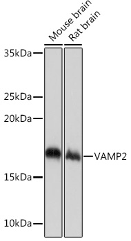
Western blot analysis of various lysates using VAMP2 Rabbit mAb (A4235) at 1:1000 dilution. Secondary antibody: HRP-conjugated Goat anti-Rabbit IgG (H+L) (AS014) at 1:10000 dilution. Lysates/proteins: 25µg per lane. Blocking buffer: 3% nonfat dry milk in TBST. Detection: ECL Basic Kit (RM00020). Exposure time: 1s. |
|
|
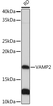
Western blot analysis of lysates from RD cells, using VAMP2 Rabbit mAb (A4235) at 1:1000 dilution. Secondary antibody: HRP-conjugated Goat anti-Rabbit IgG (H+L) (AS014) at 1:10000 dilution. Lysates/proteins: 25µg per lane. Blocking buffer: 3% nonfat dry milk in TBST. Detection: ECL Basic Kit (RM00020). Exposure time: 30s. |
|
|
Immunohistochemistry analysis of paraffin-embedded Mouse brain tissue using VAMP2 Rabbit mAb (A4235) at a dilution of 1:3000 (40x lens). High pressure antigen retrieval performed with 0.01M Tris-EDTA Buffer (pH 9.0) prior to IHC staining. |
|
|
Immunohistochemistry analysis of paraffin-embedded Human brain tissue using VAMP2 Rabbit mAb (A4235) at a dilution of 1:3000 (40x lens). High pressure antigen retrieval performed with 0.01M Tris-EDTA Buffer (pH 9.0) prior to IHC staining. |
|
|
Immunohistochemistry analysis of paraffin-embedded Mouse colon tissue using VAMP2 Rabbit mAb (A4235) at a dilution of 1:3000 (40x lens). High pressure antigen retrieval performed with 0.01M Tris-EDTA Buffer (pH 9.0) prior to IHC staining. |
|
|
Immunohistochemistry analysis of paraffin-embedded Rat brain tissue using VAMP2 Rabbit mAb (A4235) at a dilution of 1:3000 (40x lens). High pressure antigen retrieval performed with 0.01M Tris-EDTA Buffer (pH 9.0) prior to IHC staining. |
|
|
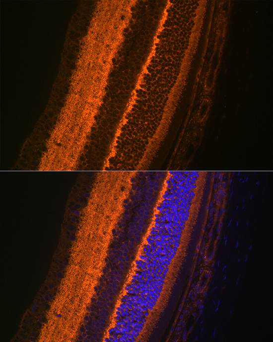
Confocal imaging ofparaffin-embedded Rat eye usingVAMP2 Rabbit mAb (A4235,dilution 1:200) followed by a further incubation with Cy3 Goat Anti-Rabbit IgG (H+L) (AS007,dilution 1:500)(Red).DAPI was used for nuclear staining (Blue). Objective: 40x. Perform high pressure antigen retrieval with 0.01 M citRate buffer (pH 6.0) prior to IF staining. |
|
|
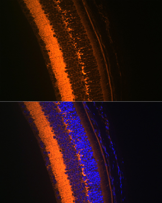
Confocal imaging ofparaffin-embedded Mouse eye usingVAMP2 Rabbit mAb (A4235,dilution 1:200) followed by a further incubation with Cy3 Goat Anti-Rabbit IgG (H+L) (AS007,dilution 1:500)(Red).DAPI was used for nuclear staining (Blue). Objective: 40x. Perform high pressure antigen retrieval with 0.01 M citRate buffer (pH 6.0) prior to IF staining. |
|
|
Confocal imaging ofparaffin-embedded Mouse brain usingVAMP2 Rabbit mAb (A4235,dilution 1:200) followed by a further incubation with Cy3 Goat Anti-Rabbit IgG (H+L) (AS007,dilution 1:500)(Red).DAPI was used for nuclear staining (Blue). Objective: 40x. Perform microwave antigen retrieval with 0.01 M citRate buffer (pH 6.0) prior to IF staining. |
Produktgarantie und fachkundiger Support
