
VAMP1 Rabbit mAb, Unconjugated, Monoclonal
Artikelnummer:
ABB-A3533
- Bilder (8)

| Artikelname: | VAMP1 Rabbit mAb, Unconjugated, Monoclonal |
| Artikelnummer: | ABB-A3533 |
| Hersteller Artikelnummer: | A3533 |
| Alternativnummer: | ABB-A3533-20UL,ABB-A3533-100UL |
| Hersteller: | ABclonal |
| Wirt: | Rabbit |
| Kategorie: | Antikörper |
| Applikation: | ELISA, IF, IHC-P, WB |
| Spezies Reaktivität: | Human |
| Immunogen: | Recombinant protein (or fragment).This information is considered to be commercially sensitive. |
| Konjugation: | Unconjugated |
| Alternative Synonym: | SAX1, SYB1, CMS25, SPAX1, VAMP-1, VAMP1 |
| Synapotobrevins, syntaxins, and the synaptosomal-associated protein SNAP25 are the main components of a protein complex involved in the docking and/or fusion of synaptic vesicles with the presynaptic membrane. The protein encoded by this gene is a member of the vesicle-associated membrane protein (VAMP)/synaptobrevin family. Mutations in this gene are associated with autosomal dominant spastic ataxia 1. Multiple alternative splice variants have been described, but the full-length nature of some variants has not been defined. |
| Application Verdünnung: | WB,1:500 - 1:2000|IHC-P,1:50 - 1:200|IF/ICC,1:50 - 1:200|ELISA,Recommended starting concentration is 1 µg/mL. Please optimize the concentration based on your specific assay requirements. |
| Anwendungsbeschreibung: | Cross-Reactivity: Human,Mouse,Rat. ResearchArea: Signal Transduction,Neuroscience, Cell Type Marker,Neuron marker,Synapse marker. Shipping: Ice Bag |
|
|
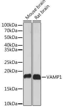
Western blot analysis of various lysates using VAMP1 Rabbit mAb (A3533) at 1:1000 dilution. Secondary antibody: HRP-conjugated Goat anti-Rabbit IgG (H+L) (AS014) at 1:10000 dilution. Lysates/proteins: 25µg per lane. Blocking buffer: 3% nonfat dry milk in TBST. Detection: ECL Basic Kit (RM00020). Exposure time: 30s. |
|
|
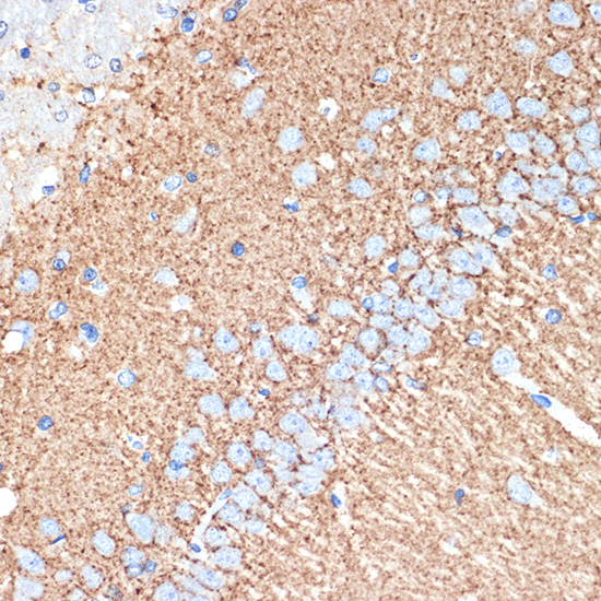
Immunohistochemistry analysis of paraffin-embeddedRat brain tissue usingVAMP1 Rabbit mAb(A3533) at a dilution of 1:200 (40x lens).High pressure antigen retrieval was performed with 0.01 M citrate buffer (pH 6.0) prior to IHC staining. |
|
|
Immunohistochemistry analysis of paraffin-embeddedMouse colon tissue usingVAMP1 Rabbit mAb(A3533) at a dilution of 1:200 (40x lens).High pressure antigen retrieval was performed with 0.01 M citrate buffer (pH 6.0) prior to IHC staining. |
|
|
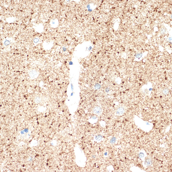
Immunohistochemistry analysis of paraffin-embeddedMouse brain tissue usingVAMP1 Rabbit mAb(A3533) at a dilution of 1:200 (40x lens).High pressure antigen retrieval was performed with 0.01 M citrate buffer (pH 6.0) prior to IHC staining. |
|
|
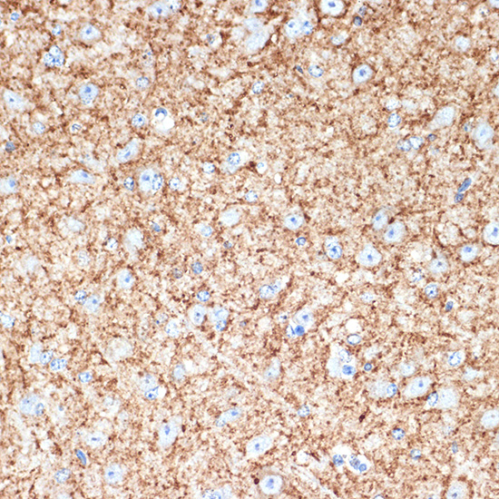
Immunohistochemistry analysis of paraffin-embeddedHuman brain tissue usingVAMP1 Rabbit mAb(A3533) at a dilution of 1:200 (40x lens).High pressure antigen retrieval was performed with 0.01 M citrate buffer (pH 6.0) prior to IHC staining. |
|
|
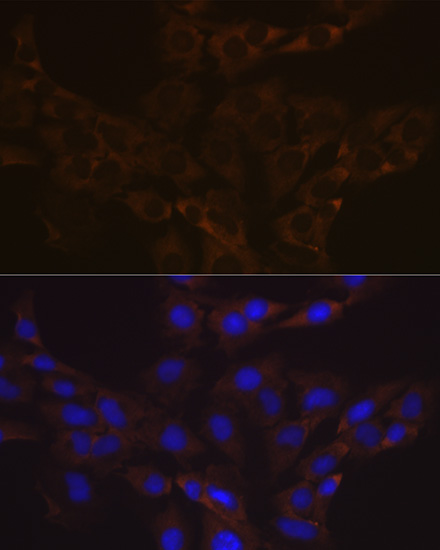
Immunofluorescence analysis of C6 cells using VAMP1 Rabbit mAb (A3533) at dilution of 1:100 (40x lens). Secondary antibody: Cy3-conjugated Goat anti-Rabbit IgG (H+L) (AS007) at 1:500 dilution. Blue: DAPI for nuclear staining. |
|
|
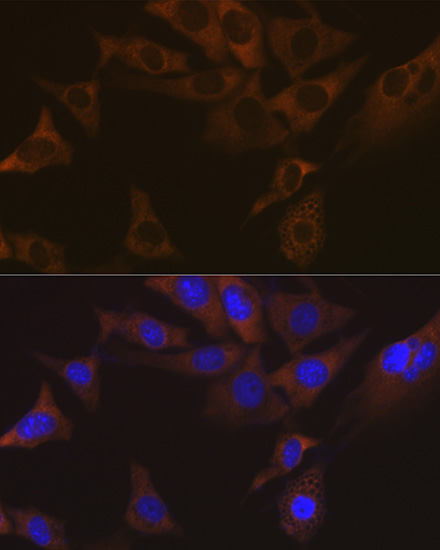
Immunofluorescence analysis of NIH-3T3 cells using VAMP1 Rabbit mAb (A3533) at dilution of 1:100 (40x lens). Secondary antibody: Cy3-conjugated Goat anti-Rabbit IgG (H+L) (AS007) at 1:500 dilution. Blue: DAPI for nuclear staining. |
|
|
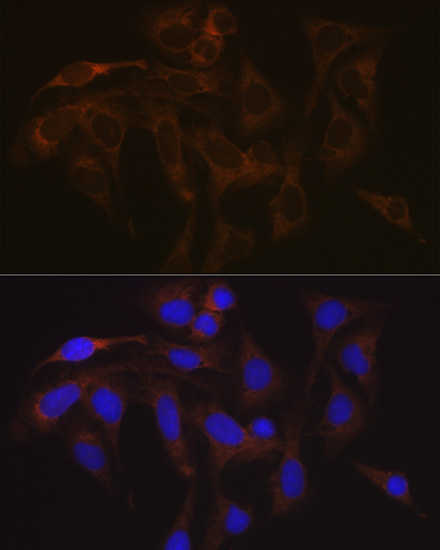
Immunofluorescence analysis of U-2 OS cells using VAMP1 Rabbit mAb (A3533) at dilution of 1:100 (40x lens). Secondary antibody: Cy3-conjugated Goat anti-Rabbit IgG (H+L) (AS007) at 1:500 dilution. Blue: DAPI for nuclear staining. |
Produktgarantie und fachkundiger Support
