
SETD2 Rabbit pAb, Unconjugated, Polyclonal
Artikelnummer:
ABB-A3194
- Bilder (8)

| Artikelname: | SETD2 Rabbit pAb, Unconjugated, Polyclonal |
| Artikelnummer: | ABB-A3194 |
| Hersteller Artikelnummer: | A3194 |
| Alternativnummer: | ABB-A3194-500UL,ABB-A3194-1000UL,ABB-A3194-100UL,ABB-A3194-20UL |
| Hersteller: | ABclonal |
| Wirt: | Rabbit |
| Kategorie: | Antikörper |
| Applikation: | ELISA, IF, IHC-P, IP, WB |
| Spezies Reaktivität: | Human |
| Immunogen: | Synthetic peptide. This information is considered to be commercially sensitive. |
| Konjugation: | Unconjugated |
| Alternative Synonym: | LLS, HYPB, SET2, HIF-1, HIP-1, KMT3A, MRD70, RAPAS, HBP231, HSPC069, p231HBP, SETD2 |
| Huntingtons disease (HD), a neurodegenerative disorder characterized by loss of striatal neurons, is caused by an expansion of a polyglutamine tract in the HD protein huntingtin. This gene encodes a protein belonging to a class of huntingtin interacting proteins characterized by WW motifs. This protein is a histone methyltransferase that is specific for lysine-36 of histone H3, and methylation of this residue is associated with active chromatin. This protein also contains a novel transcriptional activation domain and has been found associated with hyperphosphorylated RNA polymerase II. |
| Application Verdünnung: | WB,1:500 - 1:1000|IHC-P,1:500 - 1:1000|IF/ICC,1:50 - 1:200|IP,0.5µg-4µg antibody for 400µg-600µg extracts of whole cells|ELISA,Recommended starting concentration is 1 µg/mL. Please optimize the concentration based on your specific assay requirements. |
| Anwendungsbeschreibung: | Cross-Reactivity: Human,Mouse,Rat. ResearchArea: Epigenetics Nuclear Signaling,Epigenetic writers and erasers of core Histones. Shipping: Ice Bag |
|
|
Immunohistochemistry analysis of paraffin-embedded Human spleen tissue using SETD2 Rabbit pAb (A3194) at a dilution of 1:1000 (40x lens). High pressure antigen retrieval performed with 0.01M Citrate buffer (pH 6.0) prior to IHC staining. |
|
|
Immunohistochemistry analysis of paraffin-embedded Human kidney tissue using SETD2 Rabbit pAb (A3194) at a dilution of 1:1000 (40x lens). High pressure antigen retrieval performed with 0.01M Citrate buffer (pH 6.0) prior to IHC staining. |
|
|
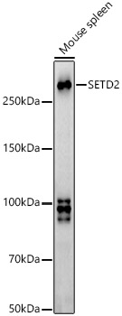
Western blot analysis of lysates from Mouse spleen, using SETD2 Rabbit pAb (A3194) at 1:900 dilution. Secondary antibody: HRP-conjugated Goat anti-Rabbit IgG (H+L) (AS014) at 1:10000 dilution. Lysates/proteins: 25µg per lane. Blocking buffer: 3% nonfat dry milk in TBST. Detection: ECL Basic Kit (RM00020). Exposure time: 90s. |
|
|
Immunohistochemistry analysis of paraffin-embedded Mouse testis tissue using SETD2 Rabbit pAb (A3194) at a dilution of 1:1000 (40x lens). High pressure antigen retrieval performed with 0.01M Citrate buffer (pH 6.0) prior to IHC staining. |
|
|
Immunohistochemistry analysis of paraffin-embedded Rat testis tissue using SETD2 Rabbit pAb (A3194) at a dilution of 1:1000 (40x lens). High pressure antigen retrieval performed with 0.01M Citrate buffer (pH 6.0) prior to IHC staining. |
|
|
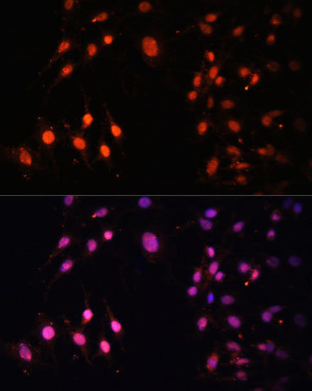
Immunofluorescence analysis of C6 cells using SETD2 Rabbit pAb (A3194) at dilution of 1:100 (40x lens). Secondary antibody: Cy3-conjugated Goat anti-Rabbit IgG (H+L) (AS007) at 1:500 dilution. Blue: DAPI for nuclear staining. |
|
|
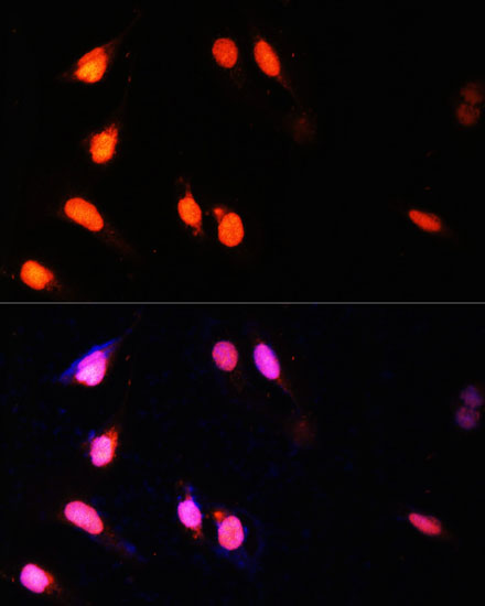
Immunofluorescence analysis of U-2 OS cells using SETD2 Rabbit pAb (A3194) at dilution of 1:100 (40x lens). Secondary antibody: Cy3-conjugated Goat anti-Rabbit IgG (H+L) (AS007) at 1:500 dilution. Blue: DAPI for nuclear staining. |
|
|
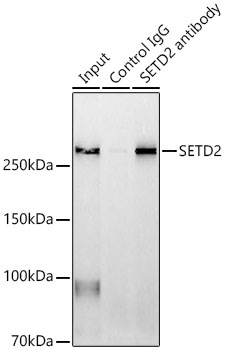
Immunoprecipitation analysis of 600 µg extracts of Mouse spleen using 3 µg SETD2 antibody (A3194). Western blot was performed from the immunoprecipitate using SETD2 antibody (A3194) at a dilution of 1:1000. |
Produktgarantie und fachkundiger Support
