
TRAP1 Rabbit pAb, Unconjugated, Polyclonal
Artikelnummer:
ABB-A2748
- Bilder (8)


| Artikelname: | TRAP1 Rabbit pAb, Unconjugated, Polyclonal |
| Artikelnummer: | ABB-A2748 |
| Hersteller Artikelnummer: | A2748 |
| Alternativnummer: | ABB-A2748-20UL,ABB-A2748-100UL,ABB-A2748-500UL,ABB-A2748-1000UL |
| Hersteller: | ABclonal |
| Wirt: | Rabbit |
| Kategorie: | Antikörper |
| Applikation: | ELISA, IF, IHC-P, WB |
| Spezies Reaktivität: | Human |
| Immunogen: | Recombinant protein (or fragment).This information is considered to be commercially sensitive. |
| Konjugation: | Unconjugated |
| Alternative Synonym: | HSP75, HSP 75, HSP90L, TRAP-1, TRAP1 |
| This gene encodes a mitochondrial chaperone protein that is member of the heat shock protein 90 (HSP90) family. The encoded protein has ATPase activity and interacts with tumor necrosis factor type I. This protein may function in regulating cellular stress responses. Alternate splicing results in multiple transcript variants. |
| Klonalität: | Polyclonal |
| Molekulargewicht: | 80kDa |
| NCBI: | 10131 |
| UniProt: | Q12931 |
| Reinheit: | Affinity purification |
| Sequenz: | GLFMREGIVTATEQEVKEDIAKLLRYESSALPSGQLTSLSEYASRMRAGTRNIYYLCAPNRHLAEHSPYYEAMKKKDTEVLFCFEQFDELTLLHLREFDKKKLISVETDIVVDHYKEEKFEDRSPAAECLSEKETEELMAWMRNVLGSRVTNVKVTLRLDTHPAMVTVLEMGAARHFLRMQQLAKTQEERAQLLQPTLEINPRHALIKKLNQLRASEPGLAQLLVDQIYENAMIAAGLVDDPRAMVGRLNELLVK |
| Target-Kategorie: | TRAP1 |
| Antibody Type: | Primary Antibody |
| Application Verdünnung: | WB,1:500 - 1:2000|IHC-P,1:50 - 1:200|IF/ICC,1:50 - 1:200|ELISA,Recommended starting concentration is 1 µg/mL. Please optimize the concentration based on your specific assay requirements. |
| Anwendungsbeschreibung: | Cross-Reactivity: Human,Mouse,Rat. ResearchArea: Epigenetics Nuclear Signaling,RNA Binding,Signal Transduction,Immunology Inflammation,Cytokines. Shipping: Ice Bag |
|
|
Western blot analysis of lysates from NIH/3T3 cells, using TRAP1 Rabbit pAb (A2748) at 1:800 dilution. Secondary antibody: HRP-conjugated Goat anti-Rabbit IgG (H+L) (AS014) at 1:10000 dilution. Lysates/proteins: 25µg per lane. Blocking buffer: 3% nonfat dry milk in TBST. Detection: ECL Basic Kit (RM00020). Exposure time: 30s. |
|
|
Immunohistochemistry analysis of paraffin-embedded Rat heart using TRAP1 Rabbit pAb (A2748) at dilution of 1:100 (40x lens). Microwave antigen retrieval performed with 0.01M PBS Buffer (pH 7.2) prior to IHC staining. |
|
|
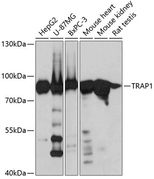
Western blot analysis of various lysates, using TRAP1 Rabbit pAb (A2748) at 1:800 dilution. Secondary antibody: HRP-conjugated Goat anti-Rabbit IgG (H+L) (AS014) at 1:10000 dilution. Lysates/proteins: 25µg per lane. Blocking buffer: 3% nonfat dry milk in TBST. Detection: ECL Basic Kit (RM00020). Exposure time: 5s. |
|
|
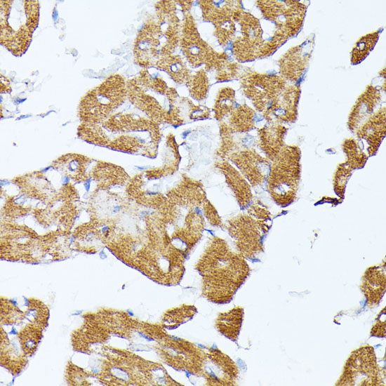
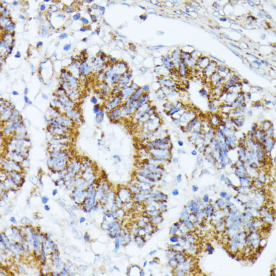
Immunohistochemistry analysis of paraffin-embedded Human colon carcinoma using TRAP1 Rabbit pAb (A2748) at dilution of 1:100 (40x lens). Microwave antigen retrieval performed with 0.01M PBS Buffer (pH 7.2) prior to IHC staining. |
|
|
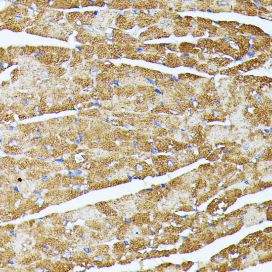
Immunohistochemistry analysis of paraffin-embedded Mouse heart using TRAP1 Rabbit pAb (A2748) at dilution of 1:100 (40x lens). Microwave antigen retrieval performed with 0.01M PBS Buffer (pH 7.2) prior to IHC staining. |
|
|
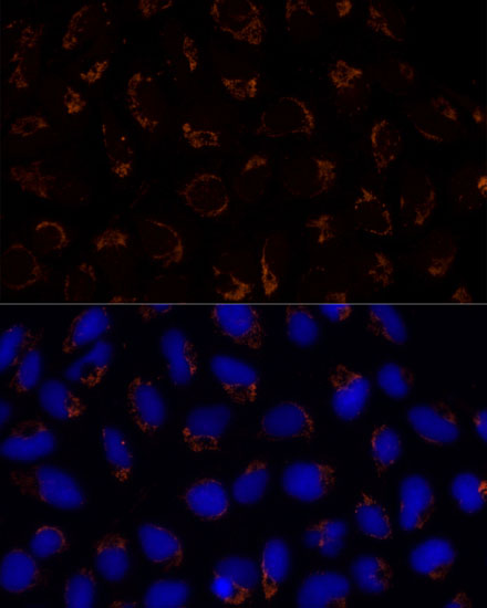
Immunofluorescence analysis of HCT 116 cells using TRAP1 Rabbit pAb (A2748) at dilution of 1:100 (40x lens). Secondary antibody: Cy3-conjugated Goat anti-Rabbit IgG (H+L) (AS007) at 1:500 dilution. Blue: DAPI for nuclear staining. |
|
|
Immunofluorescence analysis of HeLa cells using TRAP1 Rabbit pAb (A2748) at dilution of 1:100 (40x lens). Secondary antibody: Cy3-conjugated Goat anti-Rabbit IgG (H+L) (AS007) at 1:500 dilution. Blue: DAPI for nuclear staining. |
|
|
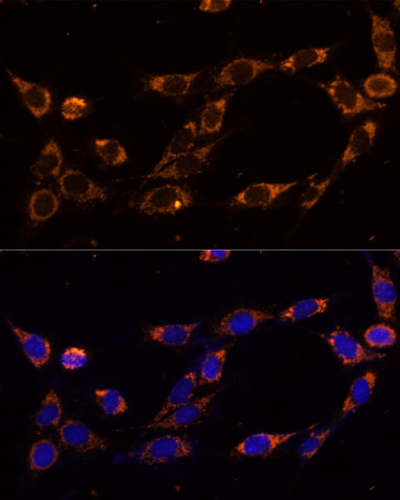
Immunofluorescence analysis of HepG2 cells using TRAP1 Rabbit pAb (A2748) at dilution of 1:100 (40x lens). Secondary antibody: Cy3-conjugated Goat anti-Rabbit IgG (H+L) (AS007) at 1:500 dilution. Blue: DAPI for nuclear staining. |
Produktgarantie und fachkundiger Support
