
IL8 Rabbit mAb, Unconjugated, Monoclonal
Artikelnummer:
ABB-A24736
- Bilder (8)

| Artikelname: | IL8 Rabbit mAb, Unconjugated, Monoclonal |
| Artikelnummer: | ABB-A24736 |
| Hersteller Artikelnummer: | A24736 |
| Alternativnummer: | ABB-A24736-100UL,ABB-A24736-20UL,ABB-A24736-500UL,ABB-A24736-1000UL |
| Hersteller: | ABclonal |
| Wirt: | Rabbit |
| Kategorie: | Antikörper |
| Applikation: | ELISA, FC, IF, IHC-P, WB |
| Spezies Reaktivität: | Human |
| Immunogen: | Recombinant protein (or fragment).This information is considered to be commercially sensitive. |
| Konjugation: | Unconjugated |
| Alternative Synonym: | IL8, NAF, GCP1, LECT, LUCT, NAP1, GCP-1, LYNAP, MDNCF, MONAP, NAP-1, SCYB8 |
| The protein encoded by this gene is a member of the CXC chemokine family and is a major mediator of the inflammatory response. The encoded protein is commonly referred to as interleukin-8 (IL-8). IL-8 is secreted by mononuclear macrophages, neutrophils, eosinophils, T lymphocytes, epithelial cells, and fibroblasts. It functions as a chemotactic factor by guiding the neutrophils to the site of infection. Bacterial and viral products rapidly induce IL-8 expression. IL-8 also participates with other cytokines in the proinflammatory signaling cascade and plays a role in systemic inflammatory response syndrome (SIRS). This gene is believed to play a role in the pathogenesis of the lower respiratory tract infection bronchiolitis, a common respiratory tract disease caused by the respiratory syncytial virus (RSV). The overproduction of this proinflammatory protein is thought to cause the lung inflammation associated with csytic fibrosis. This proinflammatory protein is also suspected of playing a role in coronary artery disease and endothelial dysfunction. This protein is also secreted by tumor cells and promotes tumor migration, invasion, angiogenesis and metastasis. This chemokine is also a potent angiogenic factor. The binding of IL-8 to one of its receptors (IL-8RB/CXCR2) increases the permeability of blood vessels and increasing levels of IL-8 are positively correlated with increased severity of multiple disease outcomes (eg, sepsis). This gene and other members of the CXC chemokine gene family form a gene cluster in a region of chromosome 4q. |
| Application Verdünnung: | WB,1:1000 - 1:6000|IHC-P,1:1000 - 1:4000|IF/ICC,1:200 - 1:400|FC (intra),1:500 - 1:1000|ELISA,Recommended starting concentration is 1 µg/mL. Please optimize the concentration based on your specific assay requirements. |
| Anwendungsbeschreibung: | Cross-Reactivity: Human. ResearchArea: Cancer,Invasion and Metastasis,Cell Biology Developmental Biology,Growth factors,Immunology Inflammation,Cytokines,Interleukins,Cell Intrinsic Innate Immunity Signaling Pathway,Cardiovascular,Angiogenesis. Shipping: Ice Bag |
|
|
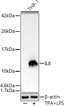
Western blot analysis of lysates from THP-1 cells using IL8 Rabbit mAb (A24736) at 1:1000 dilution. THP-1 cells were treated with PMA/TPA (80 nM) at 37°C for overnight and LPS (1 µg/ml) at 37°C for 6 hours. Secondary antibody: HRP-conjugated Goat anti-Rabbit IgG (H+L) (AS014) at 1:10000 dilution. Lysates/proteins: 25µg per lane. Blocking buffer: 3% nonfat dry milk in TBST. Detection: ECL Basic Kit (RM00020). Exposure time: 1s. |
|
|
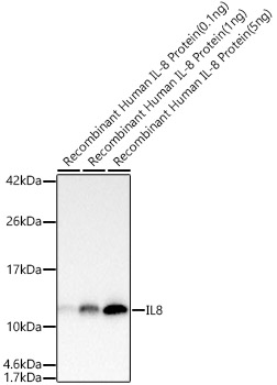
Western blot analysis of Recombinant Human IL-8/CXCL8/GCP-1 Protein (RP00052), using IL8 Rabbit mAb (A24736) at 1:1000 dilution. Secondary antibody: HRP-conjugated Goat anti-Rabbit IgG (H+L) (AS014) at 1:10000 dilution. Lysates/proteins: 5ng/1ng/0.1ng per lane. Blocking buffer: 3% nonfat dry milk in TBST. Detection: ECL Basic Kit (RM00020). Exposure time: 1s. |
|
|
Flow cytometry: 1X10 6 THP-1 cells (untreated,left) and THP-1 cells (treated with 100ng/ml TPA for 24 hours and 5 ug/mL LPS for 7 hours,right) were intracellularly-stained with IL8 Rabbit mAb (A24736,2 µg/mL,right), followed by ABflo 488 conjugated goat anti-rabbit pAb staining.. |
|
|
Immunohistochemistry analysis of paraffin-embedded Human colon carcinoma tissue using IL8 Rabbit mAb (A24736) at a dilution of 1:2000 (40x lens). High pressure antigen retrieval performed with 0.01M Tris-EDTA Buffer (pH 9.0) prior to IHC staining. |
|
|
Immunohistochemistry analysis of paraffin-embedded Human cervix cancer tissue using IL8 Rabbit mAb (A24736) at a dilution of 1:2000 (40x lens). High pressure antigen retrieval performed with 0.01M Tris-EDTA Buffer (pH 9.0) prior to IHC staining. |
|
|
Immunohistochemistry analysis of paraffin-embedded Human tonsil tissue using IL8 Rabbit mAb (A24736) at a dilution of 1:2000 (40x lens). High pressure antigen retrieval performed with 0.01M Tris-EDTA Buffer (pH 9.0) prior to IHC staining. |
|
|
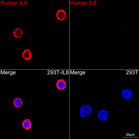
Confocal imaging of 293T cells transfected with IL8 using IL8 Rabbit mAb (A24736,dilution 1:200) followed by a further incubation with Cy3 Goat Anti-Rabbit IgG (H+L) (AS007, dilution 1:500) (Red). DAPI was used for nuclear staining (blue). Objective: 100x. |
|
|
Flow cytometry: 1X10 6 293T cells (negative control,left)and 293T (Transfection,right) cells were intracellularly-stained with IL8 Rabbit mAb (A24736,2 ug/mL,orange line) or ABflo 488 Rabbit IgG isotype control (A22069,5 µl/Test,blue line), followed by FITC conjugated goat anti-rabbit pAb staining. Non-fluorescently stained cells were used as blank control (red line). |
Produktgarantie und fachkundiger Support
