
Claudin-3 Rabbit mAb, Unconjugated, Monoclonal
Artikelnummer:
ABB-A24603
- Bilder (8)


| Artikelname: | Claudin-3 Rabbit mAb, Unconjugated, Monoclonal |
| Artikelnummer: | ABB-A24603 |
| Hersteller Artikelnummer: | A24603 |
| Alternativnummer: | ABB-A24603-20UL,ABB-A24603-100UL,ABB-A24603-500UL,ABB-A24603-1000UL |
| Hersteller: | ABclonal |
| Wirt: | Rabbit |
| Kategorie: | Antikörper |
| Applikation: | ELISA, IF, IHC-P, WB |
| Spezies Reaktivität: | Human |
| Immunogen: | Synthetic peptide. This information is considered to be commercially sensitive. |
| Konjugation: | Unconjugated |
| Alternative Synonym: | RVP1, HRVP1, C7orf1, CPE-R2, CPETR2, Claudin-3 |
| Tight junctions represent one mode of cell-to-cell adhesion in epithelial or endothelial cell sheets, forming continuous seals around cells and serving as a physical barrier to prevent solutes and water from passing freely through the paracellular space. These junctions are comprised of sets of continuous networking strands in the outwardly facing cytoplasmic leaflet, with complementary grooves in the inwardly facing extracytoplasmic leaflet. The protein encoded by this intronless gene, a member of the claudin family, is an integral membrane protein and a component of tight junction strands. It is also a low-affinity receptor for Clostridium perfringens enterotoxin, and shares aa sequence similarity with a putative apoptosis-related protein found in rat. |
| Klonalität: | Monoclonal |
| Klon-Bezeichnung: | [ARC64060] |
| Molekulargewicht: | 23kDa |
| NCBI: | 1365 |
| UniProt: | O15551 |
| Reinheit: | Affinity purification |
| Sequenz: | GVLFLLAALLTLVPVSWSANTIIRDFYNPVVPEAQKREMGAGLYVGWAAAALQLLGGALLCCSCPPREKKYTATKVVYSAPRSTGPGASLGTGYDRKDYV |
| Target-Kategorie: | CLDN3 |
| Antibody Type: | Primary Antibody |
| Application Verdünnung: | WB,1:1000 - 1:6000|IHC-P,1:200 - 1:800|IF,1:200 - 1:2000|ELISA,Recommended starting concentration is 1 µg/mL. Please optimize the concentration based on your specific assay requirements. |
| Anwendungsbeschreibung: | Cross-Reactivity: Human. ResearchArea: Cancer,Signal Transduction,Cell Biology Developmental Biology,Cell Adhesion,Tight Junctions,Cytoskeleton,Endocrine Metabolism. Shipping: Ice Bag |
|
|
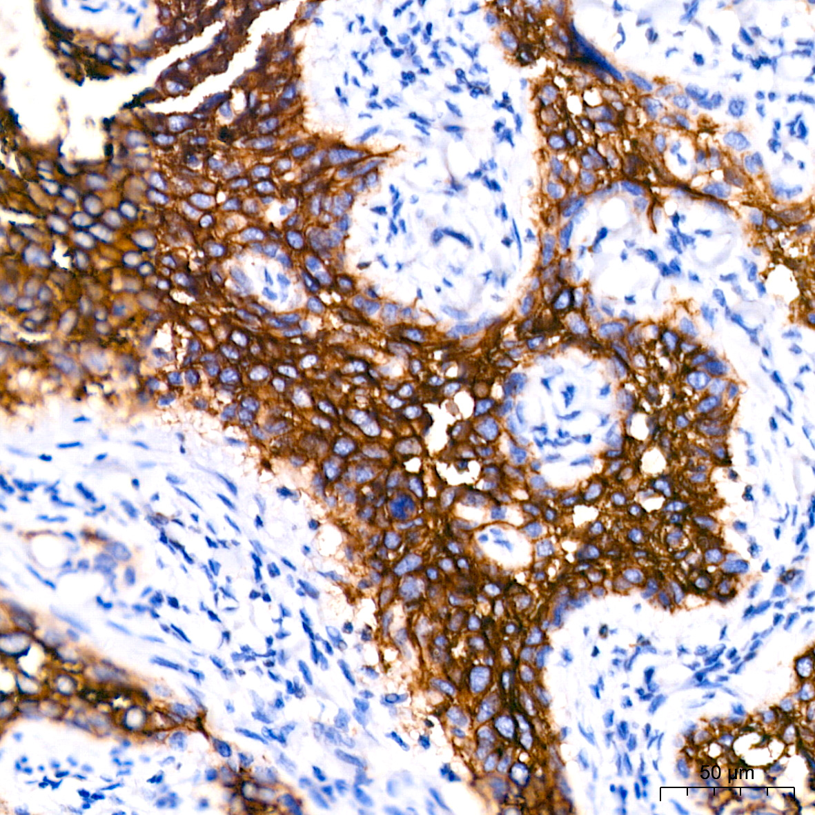
Immunohistochemistry analysis of paraffin-embedded Human lung adenocarcinoma tissue using Claudin-3 Rabbit mAb (A24603) at a dilution of 1:200 (40x lens). High pressure antigen retrieval performed with 0.01M Citrate Buffer(pH 6.0) prior to IHC staining. |
|
|
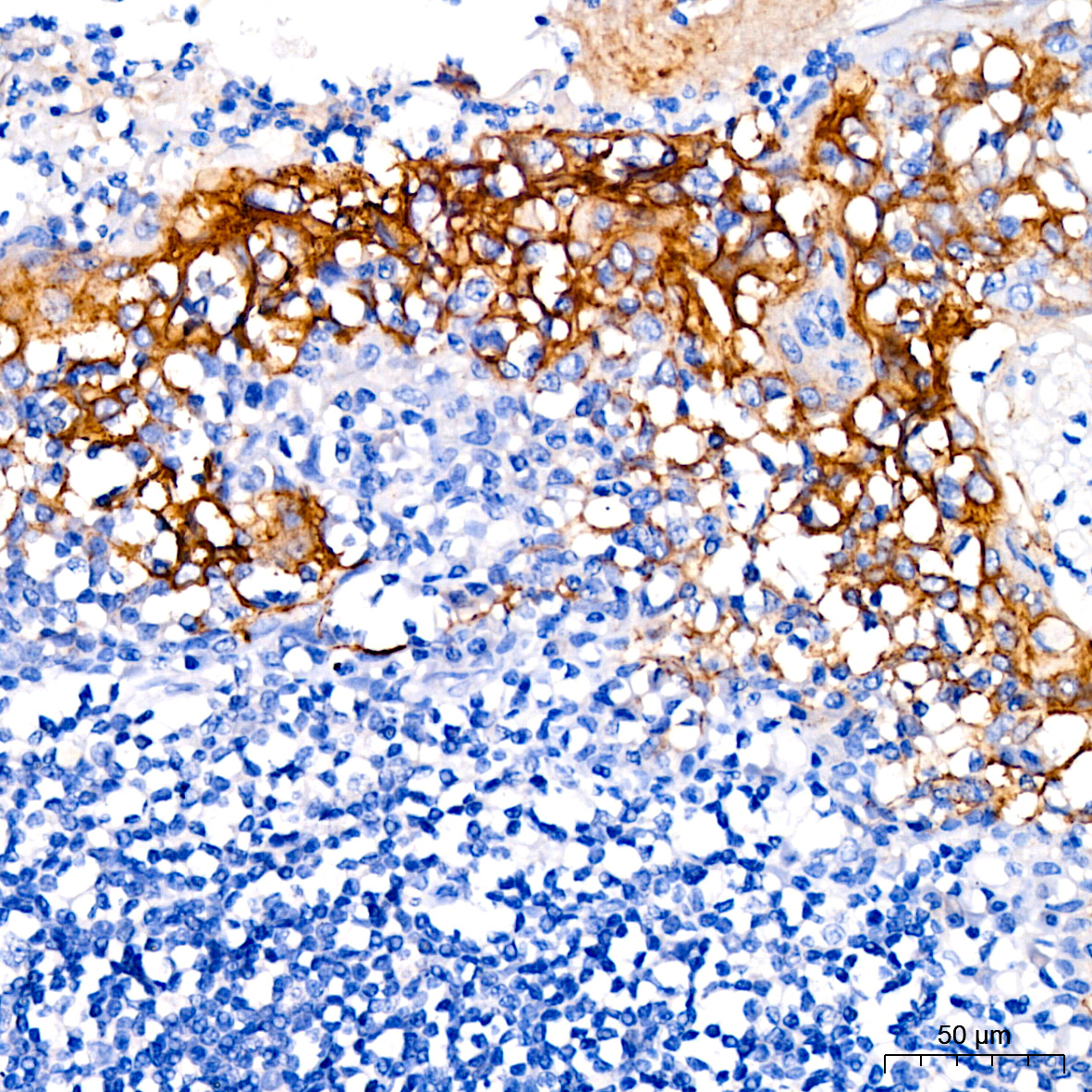
Immunohistochemistry analysis of paraffin-embedded Human colon carcinoma tissue using Claudin-3 Rabbit mAb (A24603) at a dilution of 1:200 (40x lens). High pressure antigen retrieval performed with 0.01M Citrate Buffer(pH 6.0) prior to IHC staining. |
|
|
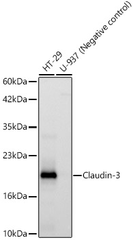
Western blot analysis of various lysates, using Claudin-3 Rabbit mAb (A24603) at 1:1000 dilution. Secondary antibody: HRP-conjugated Goat anti-Rabbit IgG (H+L) (AS014) at 1:10000 dilution. Lysates/proteins: 25µg per lane. Blocking buffer: 3% nonfat dry milk in TBST. Detection: ECL Basic Kit (RM00020). Exposure time: 10s. |
|
|
Immunohistochemistry analysis of paraffin-embedded Human oophoroma tissue using Claudin-3 Rabbit mAb (A24603) at a dilution of 1:200 (40x lens). High pressure antigen retrieval performed with 0.01M Citrate Buffer(pH 6.0) prior to IHC staining. |
|
|
Immunohistochemistry analysis of paraffin-embedded Human pancreas tissue using Claudin-3 Rabbit mAb (A24603) at a dilution of 1:200 (40x lens). High pressure antigen retrieval performed with 0.01M Citrate Buffer(pH 6.0) prior to IHC staining. |
|
|
Immunohistochemistry analysis of paraffin-embedded Human tonsil tissue using Claudin-3 Rabbit mAb (A24603) at a dilution of 1:200 (40x lens). High pressure antigen retrieval performed with 0.01M Citrate Buffer(pH 6.0) prior to IHC staining. |
|
|
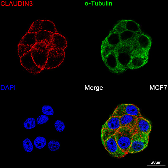
Confocal imaging of MCF7 cells using Claudin-3 Rabbit mAb (A24603,dilution 1:200)(Red). The cells were counterstained with alpha-Tubulin Mouse mAb (AC012,dilution 1:400) (Green). DAPI was used for nuclear staining (blue). Objective: 100x. |
|
|
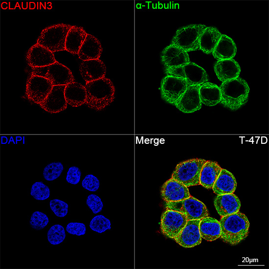
Confocal imaging of T-47D cells using Claudin-3 Rabbit mAb (A24603,dilution 1:200)(Red). The cells were counterstained with alpha-Tubulin Mouse mAb (AC012,dilution 1:400) (Green). DAPI was used for nuclear staining (blue). Objective: 100x. |
Produktgarantie und fachkundiger Support
