The protein encoded by this intronless gene is an endothelial-specific type I membrane receptor that binds thrombin. This binding results in the activation of protein C, which degrades clotting factors Va and VIIIa and reduces the amount of thrombin generated. Mutations in this gene are a cause of thromboembolic disease, also known as inherited thrombophilia.
IHC-P,1:500 - 1:1000|IF/ICC,1:50 - 1:200|FC,1:100-1:500|ELISA,Recommended starting concentration is 1 µg/mL. Please optimize the concentration based on your specific assay requirements.
Anwendungsbeschreibung:
Cross-Reactivity: Human. ResearchArea: Immunology Inflammation,CDs,Stem Cells,Cardiovascular,Blood. Shipping: Ice Bag
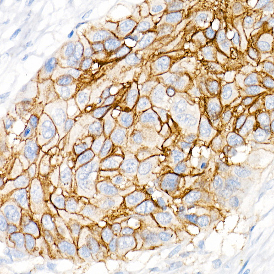
Immunohistochemistry analysis of paraffin-embedded Human lung squamous carcinoma tissue using CD141/Thrombomodulin Rabbit mAb (A21238) at dilution of 1:1000 (40x lens). High pressure antigen retrieval performed with 0.01M Citrate buffer (pH 6.0) prior to IHC staining.
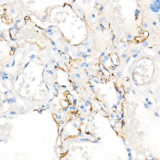
Immunohistochemistry analysis of paraffin-embedded Human lung using CD141/Thrombomodulin Rabbit mAb (A21238) at dilution of 1:1000 (40x lens). High pressure antigen retrieval performed with 0.01M Citrate buffer (pH 6.0) prior to IHC staining.
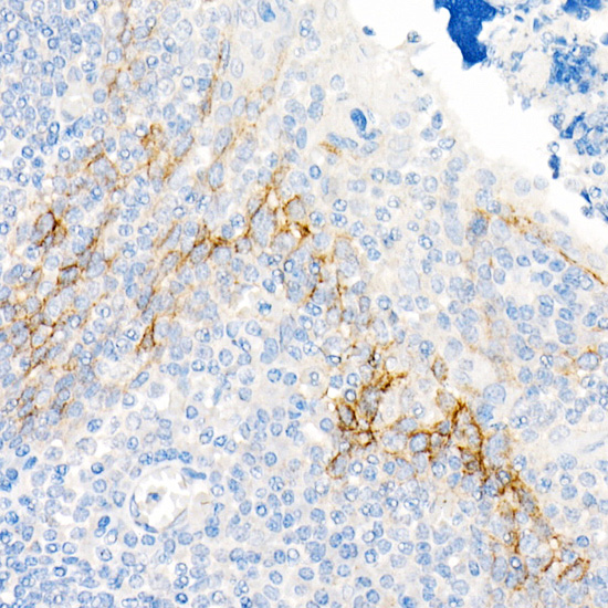
Immunohistochemistry analysis of paraffin-embedded Human tonsil using CD141/Thrombomodulin Rabbit mAb (A21238) at dilution of 1:1000 (40x lens). High pressure antigen retrieval performed with 0.01M Citrate buffer (pH 6.0) prior to IHC staining.
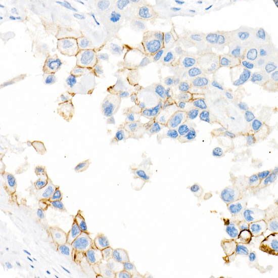
Immunohistochemistry analysis of paraffin-embedded Human urothelial carcinoma using CD141/Thrombomodulin Rabbit mAb (A21238) at dilution of 1:1000 (40x lens). High pressure antigen retrieval performed with 0.01M Citrate buffer (pH 6.0) prior to IHC staining.
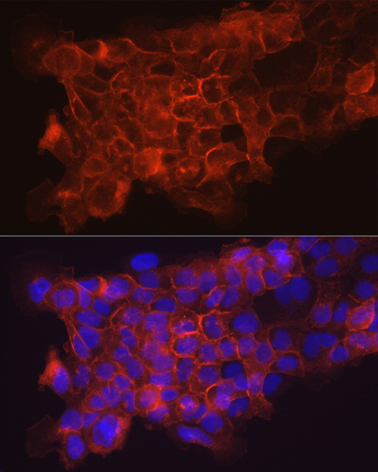
Immunofluorescence analysis of A431 cells using CD141/Thrombomodulin Rabbit mAb (A21238) at dilution of 1:100 (40x lens). Secondary antibody: Cy3-conjugated Goat anti-Rabbit IgG (H+L) (AS007) at 1:500 dilution. Blue: DAPI for nuclear staining.
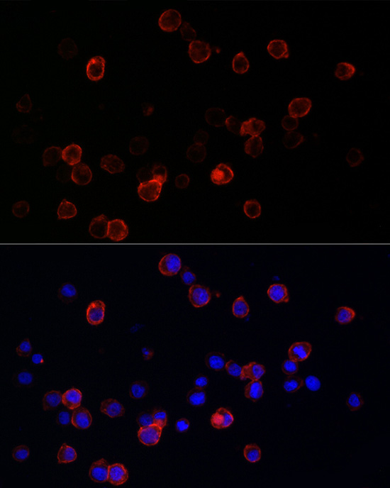
Immunofluorescence analysis of THP-1 cells using CD141/Thrombomodulin Rabbit mAb (A21238) at dilution of 1:100 (40x lens). Secondary antibody: Cy3-conjugated Goat anti-Rabbit IgG (H+L) (AS007) at 1:500 dilution. Blue: DAPI for nuclear staining.
Flow cytometry: 1X10 6 Jurkat cells (negative control,left)and THP-1 cells (right) were surface-stained with CD141/Thrombomodulin Rabbit mAb (A21238,2.5 µg/mL,orange line) or ABflo 647 Rabbit IgG isotype control (A22070,5 µl/Test,blue line), followed by Alexa Fluor 647 conjugated goat anti-rabbit pAb staining. Non-fluorescently stained cells were used as blank control (red line).
Flow cytometry: 1X10 6 THP-1 cells were surface-stained with ABflo 647 Rabbit IgG isotype control (A22070,5 µl/Test,left) or CD141/Thrombomodulin Rabbit mAb (A21238,2.5 µg/mL,right).
* Mehrwertsteuer und Versandkosten nicht enthalten. Irrtümer und Preisänderungen vorbehalten