
BMPR1B Rabbit pAb, Unconjugated, Polyclonal
Artikelnummer:
ABB-A2005
- Bilder (8)
| Artikelname: | BMPR1B Rabbit pAb, Unconjugated, Polyclonal |
| Artikelnummer: | ABB-A2005 |
| Hersteller Artikelnummer: | A2005 |
| Alternativnummer: | ABB-A2005-100UL,ABB-A2005-20UL,ABB-A2005-500UL,ABB-A2005-1000UL |
| Hersteller: | ABclonal |
| Wirt: | Rabbit |
| Kategorie: | Antikörper |
| Applikation: | ELISA, IF, IHC-P, WB |
| Spezies Reaktivität: | Human |
| Immunogen: | Recombinant protein (or fragment).This information is considered to be commercially sensitive. |
| Konjugation: | Unconjugated |
| Alternative Synonym: | ALK6, AMD3, AMDD, BDA2, ALK-6, BDA1D, CDw293, BMPR1B |
| This gene encodes a member of the bone morphogenetic protein (BMP) receptor family of transmembrane serine/threonine kinases. The ligands of this receptor are BMPs, which are members of the TGF-beta superfamily. BMPs are involved in endochondral bone formation and embryogenesis. These proteins transduce their signals through the formation of heteromeric complexes of 2 different types of serine (threonine) kinase receptors: type I receptors of about 50-55 kD and type II receptors of about 70-80 kD. Type II receptors bind ligands in the absence of type I receptors, but they require their respective type I receptors for signaling, whereas type I receptors require their respective type II receptors for ligand binding. Mutations in this gene have been associated with primary pulmonary hypertension. Several transcript variants encoding two different isoforms have been found for this gene. |
| Application Verdünnung: | WB,1:500 - 1:1000|IHC-P,1:100 - 1:500|IF/ICC,1:50 - 1:200|ELISA,Recommended starting concentration is 1 µg/mL. Please optimize the concentration based on your specific assay requirements. |
| Anwendungsbeschreibung: | Cross-Reactivity: Human,Mouse,Rat. ResearchArea: Epigenetics Nuclear Signaling,Signal Transduction,Kinase,Cell Biology Developmental Biology,Cytoskeleton,Extracellular Matrix,Bone,Growth factors,Immunology Inflammation,CDs,Stem Cells,Mesenchymal Stem Cells. Shipping: Ice Bag |
|
|
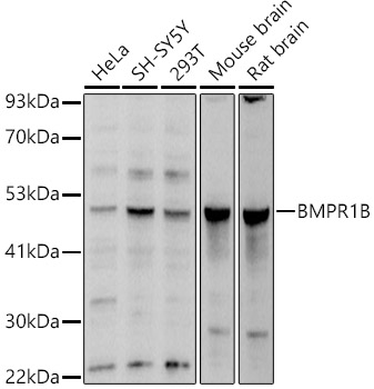
Western blot analysis of various lysates using BMPR1B Rabbit pAb (A2005) at 1:1000 dilution. Secondary antibody: HRP-conjugated Goat anti-Rabbit IgG (H+L) (AS014) at 1:10000 dilution. Lysates/proteins: 25µg per lane. Blocking buffer: 3% nonfat dry milk in TBST. Detection: ECL Basic Kit (RM00020). Exposure time: 1s. |
|
|
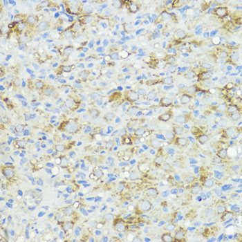
Immunohistochemistry analysis of paraffin-embedded Human mammary cancer using BMPR1B Rabbit pAb (A2005) at dilution of 1:200 (40x lens). Microwave antigen retrieval performed with 0.01M PBS Buffer (pH 7.2) prior to IHC staining. |
|
|
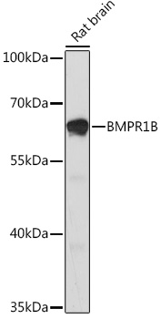
Western blot analysis of lysates from Rat brain, using BMPR1B Rabbit pAb (A2005) at 1:1000 dilution. Secondary antibody: HRP-conjugated Goat anti-Rabbit IgG (H+L) (AS014) at 1:10000 dilution. Lysates/proteins: 25µg per lane. Blocking buffer: 3% nonfat dry milk in TBST. Detection: ECL Enhanced Kit (RM00021). Exposure time: 3min. |
|
|
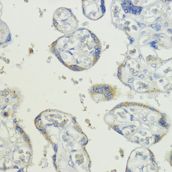
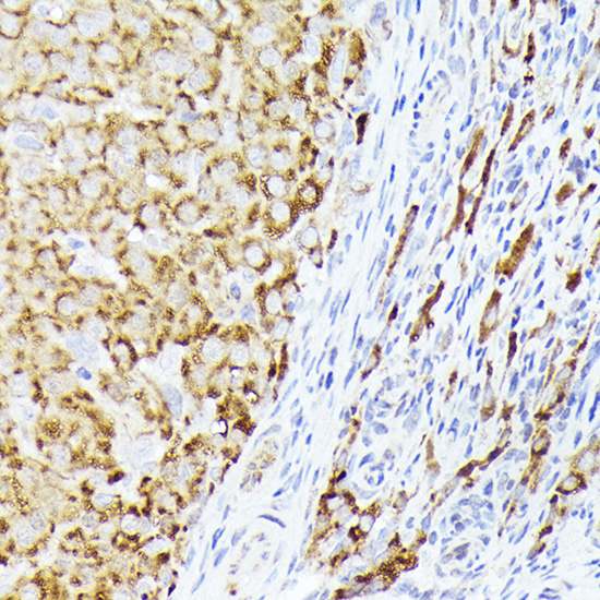
Immunohistochemistry analysis of paraffin-embedded Human placenta using BMPR1B Rabbit pAb (A2005) at dilution of 1:200 (40x lens). Microwave antigen retrieval performed with 0.01M PBS Buffer (pH 7.2) prior to IHC staining. |
|
|
Immunohistochemistry analysis of paraffin-embedded Rat ovary using BMPR1B Rabbit pAb (A2005) at dilution of 1:200 (40x lens). Microwave antigen retrieval performed with 0.01M PBS Buffer (pH 7.2) prior to IHC staining. |
|
|
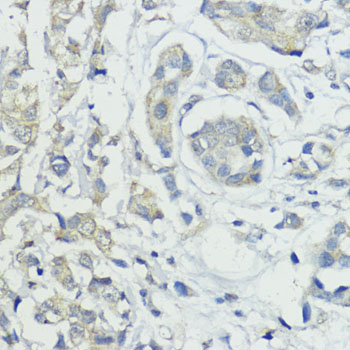
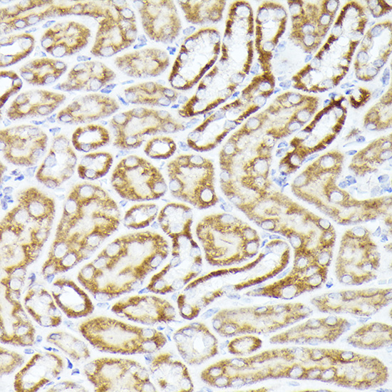
Immunohistochemistry analysis of paraffin-embedded Mouse kidney using BMPR1B Rabbit pAb (A2005) at dilution of 1:300 (40x lens). High pressure antigen retrieval performed with 0.01M Citrate buffer (pH 6.0) prior to IHC staining. |
|
|
Immunohistochemistry analysis of paraffin-embedded Rat ovary using BMPR1B Rabbit pAb (A2005) at dilution of 1:300 (40x lens). High pressure antigen retrieval performed with 0.01M Citrate buffer (pH 6.0) prior to IHC staining. |
|
|
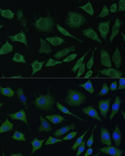
Immunofluorescence analysis of L929 cells using BMPR1B Rabbit pAb (A2005) at dilution of 1:100. Secondary antibody: Cy3-conjugated Goat anti-Rabbit IgG (H+L) (AS007) at 1:500 dilution. Blue: DAPI for nuclear staining. |
Produktgarantie und fachkundiger Support
