
[KD Validated] LC3B Rabbit mAb, Unconjugated, Monoclonal
Artikelnummer:
ABB-A19665
- Bilder (8)

| Artikelname: | [KD Validated] LC3B Rabbit mAb, Unconjugated, Monoclonal |
| Artikelnummer: | ABB-A19665 |
| Hersteller Artikelnummer: | A19665 |
| Alternativnummer: | ABB-A19665-100UL,ABB-A19665-20UL,ABB-A19665-1000UL,ABB-A19665-500UL |
| Hersteller: | ABclonal |
| Wirt: | Rabbit |
| Kategorie: | Antikörper |
| Applikation: | ELISA, IF, IHC-P, IP, WB |
| Spezies Reaktivität: | Human |
| Immunogen: | Recombinant protein (or fragment).This information is considered to be commercially sensitive. |
| Konjugation: | Unconjugated |
| Alternative Synonym: | LC3B, ATG8F, MAP1LC3B-a, MAP1A/1BLC3, 3B |
| The product of this gene is a subunit of neuronal microtubule-associated MAP1A and MAP1B proteins, which are involved in microtubule assembly and important for neurogenesis. Studies on the rat homolog implicate a role for this gene in autophagy, a process that involves the bulk degradation of cytoplasmic component. |
| Klonalität: | Monoclonal |
| Klon-Bezeichnung: | [ARC0144] |
| Molekulargewicht: | 15 kDa |
| NCBI: | 81631 |
| UniProt: | Q9GZQ8 |
| Reinheit: | Affinity purification |
| Sequenz: | PSEKTFKQRRTFEQRVEDVRLIREQHPTKIPVIIERYKGEKQLPVLDKTKFLVPDHVNMSELIKIIRRRLQLNANQAFFLLVNGHSMVSVSTPISEVYESEKDEDG |
| Target-Kategorie: | MAP1LC3B |
| Antibody Type: | Primary Antibody |
| Application Verdünnung: | WB,1:1000 - 1:4000|IHC-P,1:100 - 1:500|IF/ICC,1:200 - 1:800|IP,0.5µg-4µg antibody for 200µg-400µg extracts of whole cells|ELISA,Recommended starting concentration is 1 µg/mL. Please optimize the concentration based on your specific assay requirements. |
| Anwendungsbeschreibung: | Cross-Reactivity: Human,Mouse,Rat. ResearchArea: Cancer,Signal Transduction,Cell Biology Developmental Biology,Autophagy,Cytoskeleton,Microtubules,Endocrine Metabolism,Mitochondrial metabolism,Neuroscience,Cardiovascular,Heart. Shipping: Ice Bag |
|
|
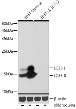
Western blot analysis of lysates from wild type(WT) and LC3B knockdown (KD) 293T cells, using [KD Validated] LC3B Rabbit mAb (A19665) at 1:1000 dilution. wild type(WT) and LC3B knockdown (KD) 293T cells were treated with Chloroquine (50 µM) at 37°C for 20 hours. Secondary antibody: HRP-conjugated Goat anti-Rabbit IgG (H+L) (AS014) at 1:10000 dilution. Lysates/proteins: 25µg per lane. Blocking buffer: 3% nonfat dry milk in TBST. Detection: ECL Basic Kit (RM00020). Exposure time: 30s. |
|
|
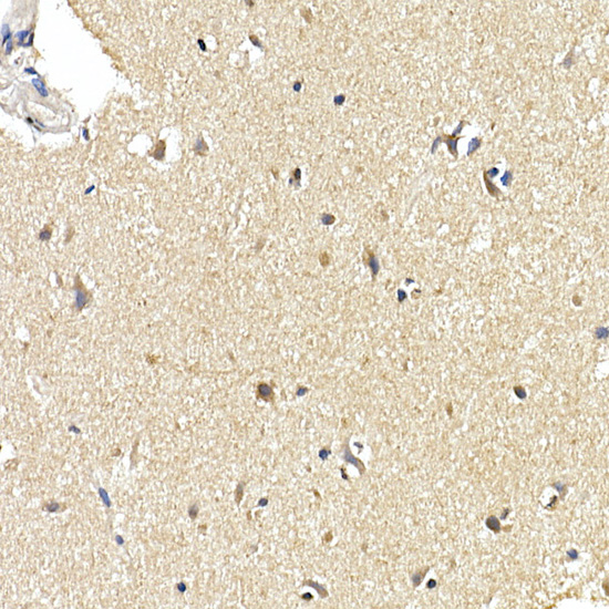
Immunohistochemistry analysis of paraffin-embedded Human brain using [KD Validated] LC3B Rabbit mAb (A19665) at dilution of 1:100 (40x lens). High pressure antigen retrieval performed with 0.01M Citrate buffer (pH 6.0) prior to IHC staining. |
|
|
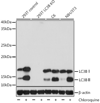
Western blot analysis of various lysates, using [KD Validated] LC3B Rabbit mAb (A19665) at 1:1000 dilution. 293T, C6 and NIH/3T3 cells were treated with Chloroquine (50 µM) at 37°C for 20 hours. Secondary antibody: HRP-conjugated Goat anti-Rabbit IgG (H+L) (AS014) at 1:10000 dilution. Lysates/proteins: 25µg per lane. Blocking buffer: 3% nonfat dry milk in TBST. Detection: ECL Basic Kit (RM00020). Exposure time: 5s. |
|
|
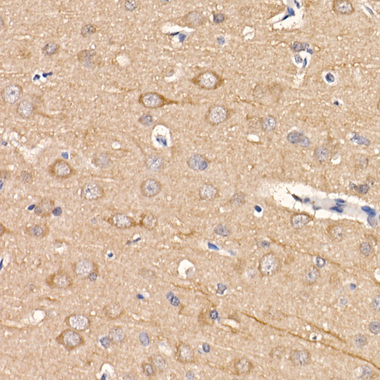
Immunohistochemistry analysis of paraffin-embedded Rat brain using [KD Validated] LC3B Rabbit mAb (A19665) at dilution of 1:100 (40x lens). High pressure antigen retrieval performed with 0.01M Citrate buffer (pH 6.0) prior to IHC staining. |
|
|
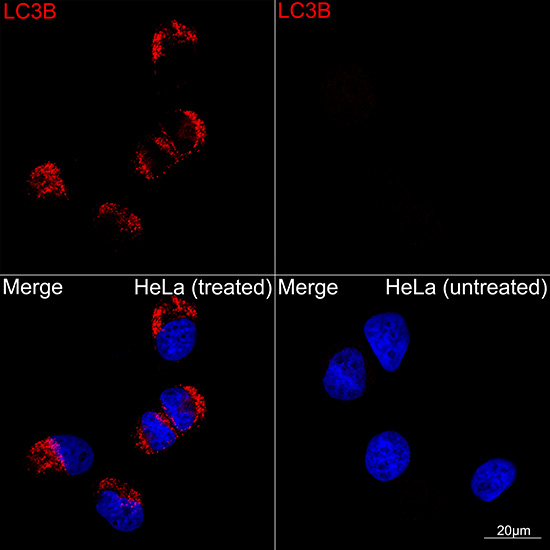
Confocal imaging of HeLa cells (treated with Chloroquine) and HeLa cells (untreated) using [KD Validated] LC3B Rabbit mAb (A19665, dilution 1:200) followed by a further incubation with Cy3 Goat Anti-Rabbit IgG (H+L) (AS007, dilution 1:500) (Red). DAPI was used for nuclear staining (Blue). Objective: 100x. |
|
|
Confocal imaging of C6 cells (treated with Chloroquine) and C6 cells (untreated) using [KD Validated] LC3B Rabbit mAb (A19665, dilution 1:200) followed by a further incubation with Cy3 Goat Anti-Rabbit IgG (H+L) (AS007, dilution 1:500) (Red). DAPI was used for nuclear staining (Blue). Objective: 100x. |
|
|
Confocal imaging of NIH/3T3 cells (treated with Chloroquine) and NIH/3T3 cells (untreated) using [KD Validated] LC3B Rabbit mAb (A19665, dilution 1:200) followed by a further incubation with Cy3 Goat Anti-Rabbit IgG (H+L) (AS007, dilution 1:500) (Red). DAPI was used for nuclear staining (Blue). Objective: 100x. |
|
|
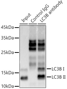
Immunoprecipitation analysis of 300 µg extracts from 293T cells using 3 µg [KD Validated] LC3B Rabbit mAb (A19665). Western blot was performed from the immunoprecipitate using LC3B antibody (A19665) at a dilution of 1:1000. |
Produktgarantie und fachkundiger Support
