
JNK1/2/3 Rabbit pAb, Polyclonal
Artikelnummer:
ABB-A18678
- Bilder (7)
| Artikelname: | JNK1/2/3 Rabbit pAb, Polyclonal |
| Artikelnummer: | ABB-A18678 |
| Hersteller Artikelnummer: | A18678 |
| Alternativnummer: | ABB-A18678-20UL,ABB-A18678-100UL,ABB-A18678-500UL,ABB-A18678-1000UL |
| Hersteller: | ABclonal |
| Wirt: | Rabbit |
| Kategorie: | Antikörper |
| Applikation: | ELISA, IF, IHC-P, IP, WB |
| Spezies Reaktivität: | Human |
| Immunogen: | Recombinant protein (or fragment).This information is considered to be commercially sensitive. |
| JNK belongs to MAP kinase family. MAP kinases act as an integration point for multiple biochemical signals, and are involved in a wide variety of cellular processes such as proliferation, differentiation, transcription regulation and development. JNK is activated by various cell stimuli, and targets specific transcription factors, and thus mediates immediate-early gene expression in response to cell stimuli. The activation of this kinase by tumor-necrosis factor alpha (TNF-alpha) is found to be required for TNF-alpha induced apoptosis. This kinase is also involved in UV radiation induced apoptosis, which is thought to be related to cytochrom c-mediated cell death pathway. Studies of the mouse counterpart of this gene suggested that this kinase play a key role in T cell proliferation, apoptosis and differentiation.JNK is expressed as ten different isoforms due to differential mRNA splicing. The predominant forms are JNK1,JNK2,and JNK3 |
| Klonalität: | Polyclonal |
| Molekulargewicht: | 35kDa/44kDa/48kDa/27kDa/52kDa |
| NCBI: | 5599 |
| UniProt: | P45983 |
| Reinheit: | Affinity purification |
| Sequenz: | MSRSKRDNNFYSVEIGDSTFTVLKRYQNLKPIGSGAQGIVCAAYDAILERNVAIKKLSRPFQNQTHAKRAYRELVLMKCVNHKNIIGLLNVFTPQKSLEEFQDVYIVMELMDANLCQVIQMELDHERMSYLLYQMLCGIKHLHSAGIIHRDLKPSNIVVKSDCTLKILDFGLARTAGTSFMMTPYVVTRYYRAPEVILGMGYKENVDLWSVGCIMGEMVCHKILFPGRDYIDQWNKVIEQLGTPCPEFMKKLQPT |
| Target-Kategorie: | MAPK8/MAPK9/MAPK10 |
| Antibody Type: | Primary Antibody |
| Application Verdünnung: | WB,1:500 - 1:2000|IHC-P,1:50 - 1:200|IF/ICC,1:50 - 1:200|IP,0.5µg-4µg antibody for 400µg-600µg extracts of whole cells|ELISA,Recommended starting concentration is 1 µg/mL. Please optimize the concentration based on your specific assay requirements. |
| Anwendungsbeschreibung: | Cross-Reactivity: Human,Mouse,Rat. Shipping: Ice Bag |
|
|
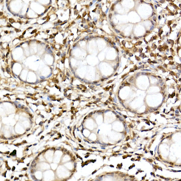
Immunohistochemistry analysis of paraffin-embedded Human colon using JNK1/2/3 Rabbit pAb (A18678) at dilution of 1:100 (40x lens). High pressure antigen retrieval performed with 0.01M Citrate buffer (pH 6.0) prior to IHC staining. |
|
|
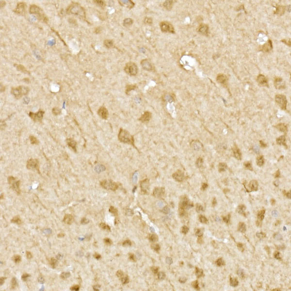
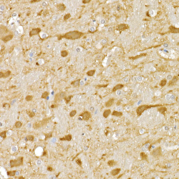
Immunohistochemistry analysis of paraffin-embedded Mouse brain using JNK1/2/3 Rabbit pAb (A18678) at dilution of 1:100 (40x lens). High pressure antigen retrieval performed with 0.01M Citrate buffer (pH 6.0) prior to IHC staining. |
|
|
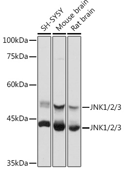
Western blot analysis of various lysates using JNK1/2/3 Rabbit pAb (A18678) at 1:1000 dilution. Secondary antibody: HRP-conjugated Goat anti-Rabbit IgG (H+L) (AS014) at 1:10000 dilution. Lysates/proteins: 25µg per lane. Blocking buffer: 3% nonfat dry milk in TBST. Detection: ECL Basic Kit (RM00020). Exposure time: 5s. |
|
|
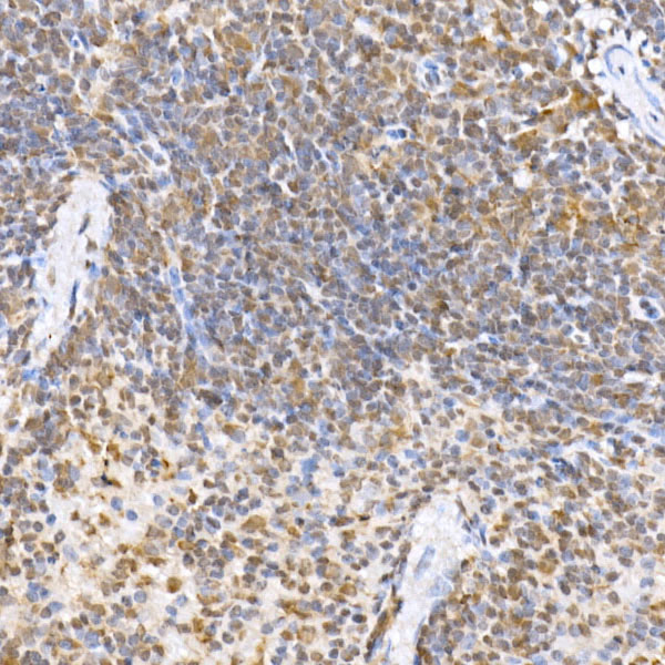
Immunohistochemistry analysis of paraffin-embedded Mouse spleen using JNK1/2/3 Rabbit pAb (A18678) at dilution of 1:100 (40x lens). High pressure antigen retrieval performed with 0.01M Citrate buffer (pH 6.0) prior to IHC staining. |
|
|
Immunohistochemistry analysis of paraffin-embedded Rat brain using JNK1/2/3 Rabbit pAb (A18678) at dilution of 1:100 (40x lens). High pressure antigen retrieval performed with 0.01M Citrate buffer (pH 6.0) prior to IHC staining. |
|
|
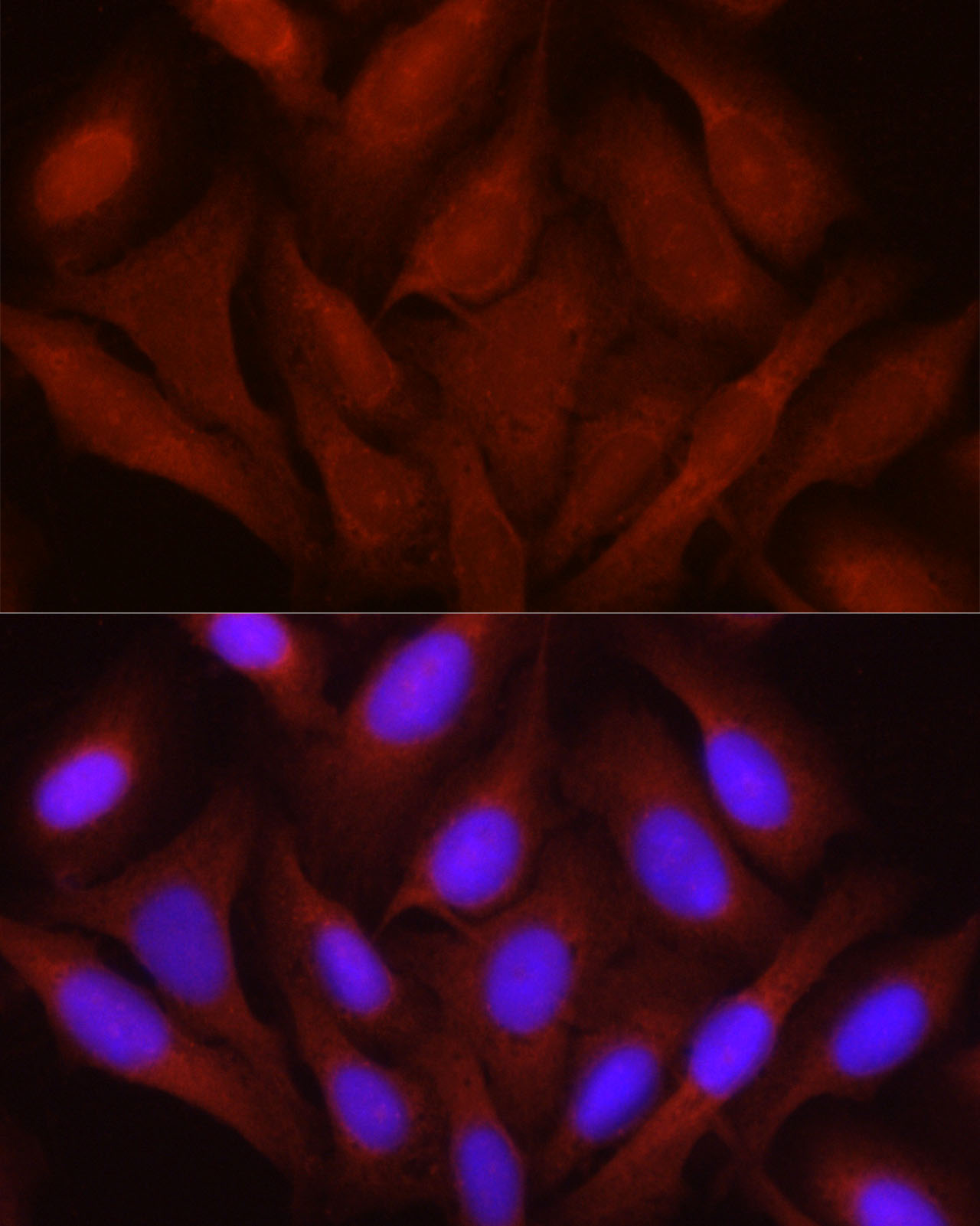
Immunofluorescence analysis of U2OS cells using JNK1/2/3 Rabbit pAb (A18678) at dilution of 1:100 (40x lens). Secondary antibody: Cy3-conjugated Goat anti-Rabbit IgG (H+L) (AS007) at 1:500 dilution. Blue: DAPI for nuclear staining. |
|
|
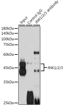
Immunoprecipitation analysis of 600 µg extracts of Mouse brain cells using 3 µg JNK1/2/3 antibody (A18678). Western blot was performed from the immunoprecipitate using JNK1/2/3 antibody (A18678) at a dilution of 1:1000. |
Produktgarantie und fachkundiger Support
