
[KD Validated] betaIII-Tubulin Rabbit mAb, Unconjugated, Monoclonal
Artikelnummer:
ABB-A17913
- Bilder (8)

| Artikelname: | [KD Validated] betaIII-Tubulin Rabbit mAb, Unconjugated, Monoclonal |
| Artikelnummer: | ABB-A17913 |
| Hersteller Artikelnummer: | A17913 |
| Alternativnummer: | ABB-A17913-100UL,ABB-A17913-20UL,ABB-A17913-500UL,ABB-A17913-1000UL |
| Hersteller: | ABclonal |
| Wirt: | Rabbit |
| Kategorie: | Antikörper |
| Applikation: | ELISA, IF, IHC-P, WB |
| Spezies Reaktivität: | Human |
| Immunogen: | Synthetic peptide. This information is considered to be commercially sensitive. |
| Konjugation: | Unconjugated |
| Alternative Synonym: | CDCBM, FEOM3, TUBB4, CDCBM1, CFEOM3, beta-4, CFEOM3A, betaIII-Tubulin |
| This gene encodes a class III member of the beta tubulin protein family. Beta tubulins are one of two core protein families (alpha and beta tubulins) that heterodimerize and assemble to form microtubules. This protein is primarily expressed in neurons and may be involved in neurogenesis and axon guidance and maintenance. Mutations in this gene are the cause of congenital fibrosis of the extraocular muscles type 3. Alternate splicing results in multiple transcript variants. A pseudogene of this gene is found on chromosome 6. |
| Klonalität: | Monoclonal |
| Klon-Bezeichnung: | [ARC0456] |
| Molekulargewicht: | 50 kDa |
| NCBI: | 10381 |
| UniProt: | Q13509 |
| Reinheit: | Affinity purification |
| Sequenz: | VAVCDIPPRGLKMSSTFIGNSTAIQELFKRISEQFTAMFRRKAFLHWYTGEGMDEMEFTEAESNMNDLVSEYQQYQDATAEEEGEMYEDDEEESEAQGPK |
| Target-Kategorie: | TUBB3 |
| Antibody Type: | Primary Antibody |
| Application Verdünnung: | WB,1:80000 - 1:400000|IF/ICC,1:200 - 1:2000|IF-F,1:200 - 1:1000|IF-P,1:200 - 1:2000|IHC-P,1:200 - 1:4000|ELISA,Recommended starting concentration is 1 µg/mL. Please optimize the concentration based on your specific assay requirements. |
| Anwendungsbeschreibung: | Cross-Reactivity: Human,Mouse,Rat. ResearchArea: Signal Transduction,Cell Biology Developmental Biology,Cell Cycle,Centrosome,Cytoskeleton,Microtubules,Neuroscience, Cell Type Marker,Neuron marker. Shipping: Ice Bag |
|
|
Western blot analysis of lysates from wild type (WT) and SRD5A2 knockdown (KD) HeLa cells using betaIII-Tubulin Rabbit mAb (A17913) at 1:200000 dilution incubated overnight at 4°C. Secondary antibody: HRP-conjugated Goat anti-Rabbit IgG (H+L) (AS014) at 1:10000 dilution. Lysates/proteins: 25 µg per lane. Blocking buffer: 3% nonfat dry milk in TBST. Detection: ECL Basic Kit (RM00020). Exposure time: 30 s. |
|
|
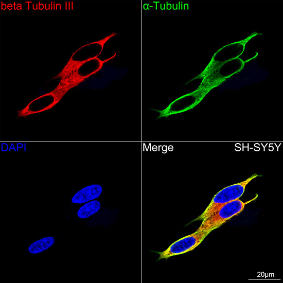
Confocal imaging of SH-SY5Y cells using betaIII-Tubulin Rabbit mAb (A17913, dilution 1:200) followed by a further incubation with Cy3 Goat Anti-Rabbit IgG (H+L) (AS007, dilution 1:500) (Red). The cells were counterstained with alpha-Tubulin Mouse mAb (AC012, dilution 1:400) followed by incubation with ABflo 488-conjugated Goat Anti-Mouse IgG (H+L) Ab (AS076, dilution 1:500) (Green). DAPI was used for nuclear staining (Blue). Objective: 100x. |
|
|
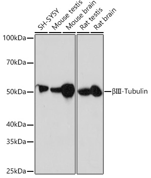
Western blot analysis of lysates from Mouse testis using betaIII-Tubulin Rabbit mAb (A17913) at 1:200000 dilution incubated overnight at 4°C. Secondary antibody: HRP-conjugated Goat anti-Rabbit IgG (H+L) (AS014) at 1:10000 dilution. Lysates/proteins: 25 µg per lane. Blocking buffer: 3% nonfat dry milk in TBST. Detection: ECL Basic Kit (RM00020). Exposure time: 1 s. |
|
|
Immunohistochemistry analysis of paraffin-embedded Mouse brain tissue using betaIII-Tubulin Rabbit mAb (A17913) at a dilution of 1:200 (40x lens). High pressure antigen retrieval performed with 0.01M Citrate buffer (pH 6.0) prior to IHC staining. |
|
|
Immunohistochemistry analysis of paraffin-embedded Rat brain tissue using betaIII-Tubulin Rabbit mAb (A17913) at a dilution of 1:200 (40x lens). High pressure antigen retrieval performed with 0.01M Citrate buffer (pH 6.0) prior to IHC staining. |
|
|
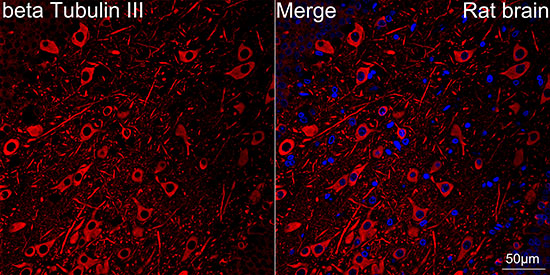
Confocal imaging of paraffin-embedded Rat brain tissue using betaIII-Tubulin Rabbit mAb (A17913, dilution 1:200) followed by a further incubation with Cy3 Goat Anti-Rabbit IgG (H+L) (AS007, dilution 1:500) (Red). DAPI was used for nuclear staining (Blue). Objective: 40x. Perform microwave antigen retrieval with 0.01M citrate buffer (pH 6.0) prior to IF staining. |
|
|
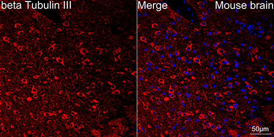
Confocal imaging of paraffin-embedded Mouse brain tissue using betaIII-Tubulin Rabbit mAb (A17913, dilution 1:200) followed by a further incubation with Cy3 Goat Anti-Rabbit IgG (H+L) (AS007, dilution 1:500) (Red). DAPI was used for nuclear staining (Blue). Objective: 40x. Perform high pressure antigen retrieval with 0.01M citrate buffer (pH 6.0) prior to IF staining. |
|
|
Confocal imaging of frozen sections Mouse brain tissue using betaIII-Tubulin Rabbit mAb (A17913, dilution 1:200) followed by a further incubation with Cy3-conjugated Goat anti-Rabbit IgG (H+L) (AS007, dilution 1:500) (Red). DAPI was used for nuclear staining (Blue). Microwave antigen retrieval performed with 0.01M Citrate Buffer (pH 6.0) prior to IF staining. Objective: 40x. |
Produktgarantie und fachkundiger Support
