
BOK Rabbit pAb, Unconjugated, Polyclonal
Artikelnummer:
ABB-A16354
- Bilder (7)

| Artikelname: | BOK Rabbit pAb, Unconjugated, Polyclonal |
| Artikelnummer: | ABB-A16354 |
| Hersteller Artikelnummer: | A16354 |
| Alternativnummer: | ABB-A16354-100UL,ABB-A16354-20UL,ABB-A16354-500UL,ABB-A16354-1000UL |
| Hersteller: | ABclonal |
| Wirt: | Rabbit |
| Kategorie: | Antikörper |
| Applikation: | ELISA, IF, IHC-P, WB |
| Spezies Reaktivität: | Human |
| Immunogen: | Synthetic peptide. This information is considered to be commercially sensitive. |
| Konjugation: | Unconjugated |
| Alternative Synonym: | BOKL, BCL2L9, BOK |
| The protein encoded by this gene belongs to the BCL2 family, members of which form homo- or heterodimers, and act as anti- or proapoptotic regulators that are involved in a wide variety of cellular processes. Studies in rat show that this protein has restricted expression in reproductive tissues, interacts strongly with some antiapoptotic BCL2 proteins, not at all with proapoptotic BCL2 proteins, and induces apoptosis in transfected cells. Thus, this protein represents a proapoptotic member of the BCL2 family. |
| Application Verdünnung: | WB,1:500 - 1:10000|IF/ICC,1:50 - 1:200|IHC-P,1:50 - 1:200|ELISA,Recommended starting concentration is 1 µg/mL. Please optimize the concentration based on your specific assay requirements. |
| Anwendungsbeschreibung: | Cross-Reactivity: Human,Mouse,Rat. ResearchArea: Cancer,Tumor suppressors,p53 pathway,Cell Biology Developmental Biology,Apoptosis. Shipping: Ice Bag |
|
|
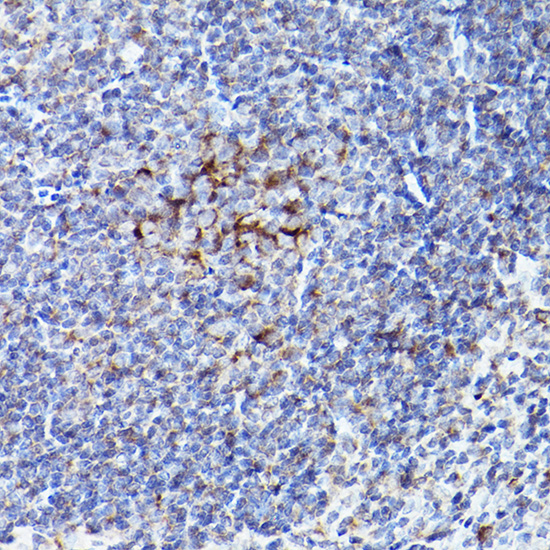
Immunohistochemistry analysis of paraffin-embedded Mouse spleen using BOK Rabbit pAb (A16354) at dilution of 1:100 (40x lens). High pressure antigen retrieval performed with 0.01M Citrate buffer (pH 6.0) prior to IHC staining. |
|
|
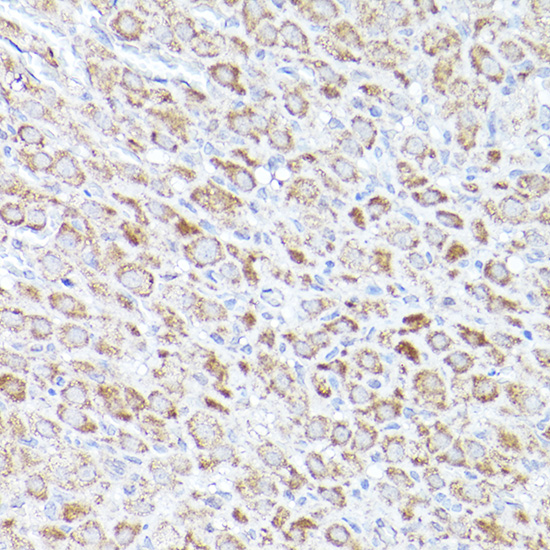
Immunohistochemistry analysis of paraffin-embedded Rat lung using BOK Rabbit pAb (A16354) at dilution of 1:100 (40x lens). High pressure antigen retrieval performed with 0.01M Citrate buffer (pH 6.0) prior to IHC staining. |
|
|
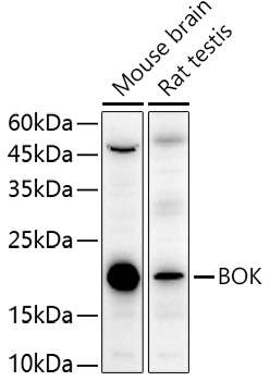
Western blot analysis of various lysates using BOK Rabbit pAb (A16354)at 1:10000 dilutionincubated overnight at 4°C. Secondary antibody: HRP-conjugated Goat anti-Rabbit IgG (H+L) (AS014) at 1:10000 dilution. Lysates/proteins: 25 µg per lane. Blocking buffer: 3% nonfat dry milk in TBST. Detection: ECL Basic Kit (RM00020). Negative control (NC): U-937. Exposure time: 90 s. |
|
|
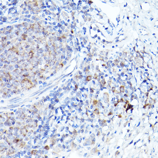
Immunohistochemistry analysis of paraffin-embedded Rat ovary using BOK Rabbit pAb (A16354) at dilution of 1:100 (40x lens). High pressure antigen retrieval performed with 0.01M Citrate buffer (pH 6.0) prior to IHC staining. |
|
|
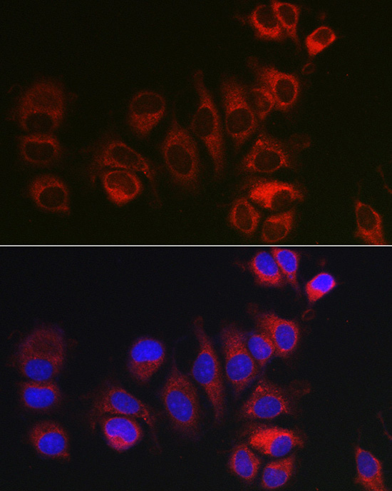
Immunofluorescence analysis of MCF7 cells using BOK Rabbit pAb (A16354) at dilution of 1:100 (40x lens). Secondary antibody: Cy3-conjugated Goat anti-Rabbit IgG (H+L) (AS007) at 1:500 dilution. Blue: DAPI for nuclear staining. |
|
|
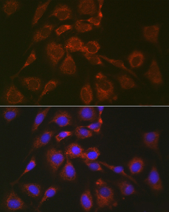
Immunofluorescence analysis of NIH/3T3 cells using BOK Rabbit pAb (A16354) at dilution of 1:100 (40x lens). Secondary antibody: Cy3-conjugated Goat anti-Rabbit IgG (H+L) (AS007) at 1:500 dilution. Blue: DAPI for nuclear staining. |
|
|
Immunofluorescence analysis of NIH/3T3 cells using BOK Rabbit pAb (A16354) at dilution of 1:100 (40x lens). Secondary antibody: Cy3-conjugated Goat anti-Rabbit IgG (H+L) (AS007) at 1:500 dilution. Blue: DAPI for nuclear staining. |
Produktgarantie und fachkundiger Support
