
IL23R Rabbit pAb, Unconjugated, Polyclonal
Artikelnummer:
ABB-A1613
- Bilder (7)

| Artikelname: | IL23R Rabbit pAb, Unconjugated, Polyclonal |
| Artikelnummer: | ABB-A1613 |
| Hersteller Artikelnummer: | A1613 |
| Alternativnummer: | ABB-A1613-100UL,ABB-A1613-20UL,ABB-A1613-1000UL,ABB-A1613-500UL |
| Hersteller: | ABclonal |
| Wirt: | Rabbit |
| Kategorie: | Antikörper |
| Applikation: | ELISA, IF, IHC-P, WB |
| Spezies Reaktivität: | Human |
| Immunogen: | Recombinant protein (or fragment).This information is considered to be commercially sensitive. |
| Konjugation: | Unconjugated |
| Alternative Synonym: | PSORS7, IL23R |
| The protein encoded by this gene is a subunit of the receptor for IL23A/IL23. This protein pairs with the receptor molecule IL12RB1/IL12Rbeta1, and both are required for IL23A signaling. This protein associates constitutively with Janus kinase 2 (JAK2), and also binds to transcription activator STAT3 in a ligand-dependent manner. |
| Application Verdünnung: | WB,1:500 - 1:5000|IF/ICC,1:50 - 1:100|IHC-P,1:50 - 1:200|ELISA,Recommended starting concentration is 1 µg/mL. Please optimize the concentration based on your specific assay requirements. |
| Anwendungsbeschreibung: | Cross-Reactivity: Human,Mouse,Rat. ResearchArea: Epigenetics Nuclear Signaling,Immunology Inflammation,Cytokines,Interleukins,Cell Intrinsic Innate Immunity Signaling Pathway. Shipping: Ice Bag |
|
|
Western blot analysis of lysates from RAW 264.7 cells using IL23R Rabbit pAb (A1613) at 1:500 dilution incubated overnight at 4°C. Secondary antibody: HRP-conjugated Goat anti-Rabbit IgG (H+L) (AS014) at 1:10000 dilution. Lysates/proteins: 25 µg per lane. Blocking buffer: 3% nonfat dry milk in TBST. Detection: ECL Basic Kit (RM00020). Exposure time: 90s. |
|
|
Western blot analysis of various lysates using IL23R Rabbit pAb (A1613) at 1:500 dilution incubated overnight at 4°C. Secondary antibody: HRP-conjugated Goat anti-Rabbit IgG (H+L) (AS014) at 1:10000 dilution. Lysates/proteins: 25 µg per lane. Blocking buffer: 3% nonfat dry milk in TBST. Detection: ECL Basic Kit (RM00020). Exposure time: 3s. |
|
|
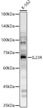
Western blot analysis of lysates from K-562 cells, using IL23R Rabbit pAb (A1613) at 1:1000 dilution. Secondary antibody: HRP-conjugated Goat anti-Rabbit IgG (H+L) (AS014) at 1:10000 dilution. Lysates/proteins: 25µg per lane. Blocking buffer: 3% nonfat dry milk in TBST. Detection: ECL Basic Kit (RM00020). Exposure time: 30s. |
|
|
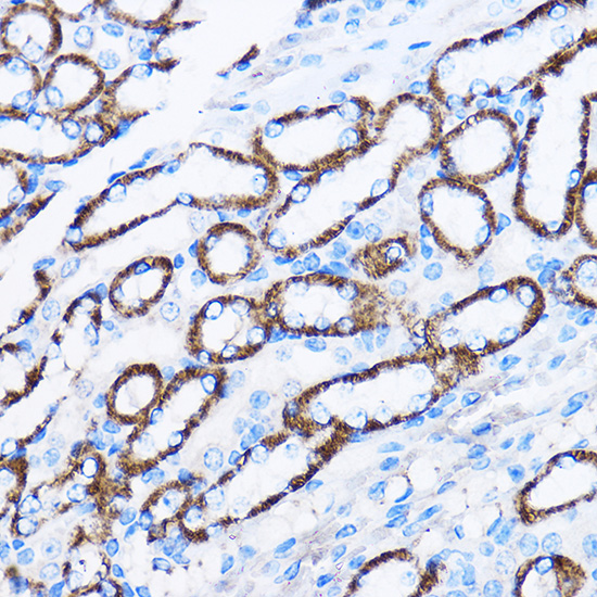
Immunohistochemistry analysis of paraffin-embedded Mouse kidney using IL23R Rabbit pAb (A1613) at dilution of 1:100 (40x lens). Microwave antigen retrieval performed with 0.01M PBS Buffer (pH 7.2) prior to IHC staining. |
|
|
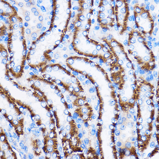
Immunohistochemistry analysis of paraffin-embedded Rat kidney using IL23R Rabbit pAb (A1613) at dilution of 1:100 (40x lens). Microwave antigen retrieval performed with 0.01M PBS Buffer (pH 7.2) prior to IHC staining. |
|
|
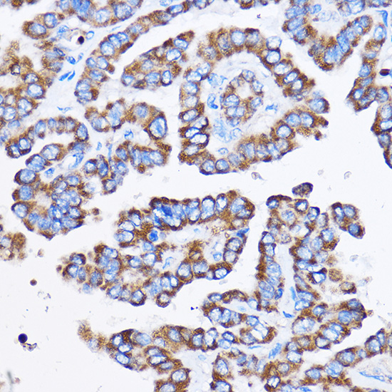
Immunohistochemistry analysis of paraffin-embedded Human thyroid cancer using IL23R Rabbit pAb (A1613) at dilution of 1:100 (40x lens). Microwave antigen retrieval performed with 0.01M PBS Buffer (pH 7.2) prior to IHC staining. |
|
|
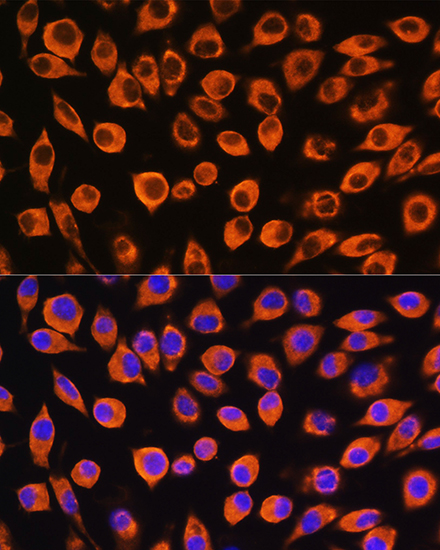
Immunofluorescence analysis of L929 cells using IL23R Rabbit pAb (A1613) at dilution of 1:100. Secondary antibody: Cy3-conjugated Goat anti-Rabbit IgG (H+L) (AS007) at 1:500 dilution. Blue: DAPI for nuclear staining. |
Produktgarantie und fachkundiger Support
