
DAPK3 Rabbit pAb, Unconjugated, Polyclonal
Artikelnummer:
ABB-A15047
- Bilder (7)


| Artikelname: | DAPK3 Rabbit pAb, Unconjugated, Polyclonal |
| Artikelnummer: | ABB-A15047 |
| Hersteller Artikelnummer: | A15047 |
| Alternativnummer: | ABB-A15047-20UL,ABB-A15047-100UL,ABB-A15047-500UL,ABB-A15047-1000UL |
| Hersteller: | ABclonal |
| Wirt: | Rabbit |
| Kategorie: | Antikörper |
| Applikation: | ELISA, IF, IHC-P, WB |
| Spezies Reaktivität: | Human |
| Immunogen: | Synthetic peptide. This information is considered to be commercially sensitive. |
| Konjugation: | Unconjugated |
| Alternative Synonym: | DLK, ZIP, ZIPK, DAPK3 |
| Death-associated protein kinase 3 (DAPK3) induces morphological changes in apoptosis when overexpressed in mammalian cells. These results suggest that DAPK3 may play a role in the induction of apoptosis. |
| Application Verdünnung: | WB,1:500 - 1:2000|IHC-P,1:50 - 1:100|IF/ICC,1:50 - 1:100|ELISA,Recommended starting concentration is 1 µg/mL. Please optimize the concentration based on your specific assay requirements. |
| Anwendungsbeschreibung: | Cross-Reactivity: Human,Mouse,Rat. ResearchArea: Cancer,Invasion and Metastasis,Signal Transduction,Kinase,Serine threonine kinases,Cell Biology Developmental Biology,Apoptosis,Death Receptor Signaling Pathway. Shipping: Ice Bag |
|
|
Immunohistochemistry analysis of paraffin-embedded Rat kidney using DAPK3 Rabbit pAb (A15047) at dilution of 1:100 (40x lens). Microwave antigen retrieval performed with 0.01M PBS Buffer (pH 7.2) prior to IHC staining. |
|
|
Immunohistochemistry analysis of paraffin-embedded Human lung cancer using DAPK3 Rabbit pAb (A15047) at dilution of 1:100 (40x lens). Microwave antigen retrieval performed with 0.01M PBS Buffer (pH 7.2) prior to IHC staining. |
|
|
Western blot analysis of various lysates using DAPK3 Rabbit pAb (A15047) at 1:1000 dilution. Secondary antibody: HRP-conjugated Goat anti-Rabbit IgG (H+L) (AS014) at 1:10000 dilution. Lysates/proteins: 25µg per lane. Blocking buffer: 3% nonfat dry milk in TBST. Detection: ECL Basic Kit (RM00020). Exposure time: 90s. |
|
|
Immunohistochemistry analysis of paraffin-embedded Human placenta using DAPK3 Rabbit pAb (A15047) at dilution of 1:100 (40x lens). Microwave antigen retrieval performed with 0.01M PBS Buffer (pH 7.2) prior to IHC staining. |
|
|
Immunohistochemistry analysis of paraffin-embedded Mouse kidney using DAPK3 Rabbit pAb (A15047) at dilution of 1:100 (40x lens). Microwave antigen retrieval performed with 0.01M PBS Buffer (pH 7.2) prior to IHC staining. |
|
|
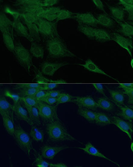
Immunofluorescence analysis of C6 cells using DAPK3 Rabbit pAb (A15047) at dilution of 1:100 (40x lens). Secondary antibody: Cy3-conjugated Goat anti-Rabbit IgG (H+L) (AS007) at 1:500 dilution. Blue: DAPI for nuclear staining. |
|
|
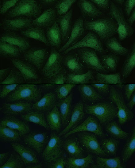
Immunofluorescence analysis of U-2 OS cells using DAPK3 Rabbit pAb (A15047) at dilution of 1:100 (40x lens). Secondary antibody: Cy3-conjugated Goat anti-Rabbit IgG (H+L) (AS007) at 1:500 dilution. Blue: DAPI for nuclear staining. |
Produktgarantie und fachkundiger Support
