
AKR1C2 Rabbit pAb, Unconjugated, Polyclonal
Artikelnummer:
ABB-A1048
- Bilder (7)


| Artikelname: | AKR1C2 Rabbit pAb, Unconjugated, Polyclonal |
| Artikelnummer: | ABB-A1048 |
| Hersteller Artikelnummer: | A1048 |
| Alternativnummer: | ABB-A1048-100UL,ABB-A1048-20UL,ABB-A1048-1000UL,ABB-A1048-500UL |
| Hersteller: | ABclonal |
| Wirt: | Rabbit |
| Kategorie: | Antikörper |
| Applikation: | ELISA, IF, IHC-P, WB |
| Spezies Reaktivität: | Human |
| Immunogen: | Recombinant protein (or fragment).This information is considered to be commercially sensitive. |
| Konjugation: | Unconjugated |
| Alternative Synonym: | DD, DD2, TDD, BABP, DD-2, DDH2, HBAB, HAKRD, MCDR2, SRXY8, DD/BABP, AKR1C-pseudo, AKR1C2 |
| This gene encodes a member of the aldo/keto reductase superfamily, which consists of more than 40 known enzymes and proteins. These enzymes catalyze the conversion of aldehydes and ketones to their corresponding alcohols using NADH and/or NADPH as cofactors. The enzymes display overlapping but distinct substrate specificity. This enzyme binds bile acid with high affinity, and shows minimal 3-alpha-hydroxysteroid dehydrogenase activity. This gene shares high sequence identity with three other gene members and is clustered with those three genes at chromosome 10p15-p14. Three transcript variants encoding two different isoforms have been found for this gene. |
| Klonalität: | Polyclonal |
| Molekulargewicht: | 37kDa |
| NCBI: | 1646 |
| UniProt: | P52895 |
| Reinheit: | Affinity purification |
| Sequenz: | MDSKYQCVKLNDGHFMPVLGFGTYAPAEVPKSKALEAVKLAIEAGFHHIDSAHVYNNEEQVGLAIRSKIADGSVKREDIFYTSKLWSNSHRPELVRPALERSLKNLQLDYVDLYLIHFPVSVKPGEEVIPKDENGKILFDTVDLCATWEAMEKCKDAGLAKSIGVSNFNHRLLEMILNKPGLKYKPVCNQVECHPYFNQRKLLDFCKSKDIVLVAYSALGSHREEPWVDPNSPVLLEDPVLCALAKKHKRTPALI |
| Target-Kategorie: | AKR1C2 |
| Antibody Type: | Primary Antibody |
| Application Verdünnung: | WB,1:1000 - 1:5000|IHC-P,1:50 - 1:200|IF/ICC,1:50 - 1:100|ELISA,Recommended starting concentration is 1 µg/mL. Please optimize the concentration based on your specific assay requirements. |
| Anwendungsbeschreibung: | Cross-Reactivity: Human,Mouse,Rat. Shipping: Ice Bag |
|
|
Immunohistochemistry analysis of paraffin-embedded Rat liver using AKR1C2 Rabbit pAb (A1048) at dilution of 1:100 (40x lens). Microwave antigen retrieval performed with 0.01M PBS Buffer (pH 7.2) prior to IHC staining. |
|
|
Immunohistochemistry analysis of paraffin-embedded Human liver cancer using AKR1C2 Rabbit pAb (A1048) at dilution of 1:100 (40x lens). Microwave antigen retrieval performed with 0.01M PBS Buffer (pH 7.2) prior to IHC staining. |
|
|
Western blot analysis of various lysates, using AKR1C2 Rabbit pAb (A1048) at 1:2000 dilution. Secondary antibody: HRP-conjugated Goat anti-Rabbit IgG (H+L) (AS014) at 1:10000 dilution. Lysates/proteins: 25µg per lane. Blocking buffer: 3% nonfat dry milk in TBST. Detection: ECL Basic Kit (RM00020). Exposure time: 10s. |
|
|
Immunohistochemistry analysis of paraffin-embedded Human Colon cancer using AKR1C2 Rabbit pAb (A1048) at dilution of 1:100 (40x lens). Microwave antigen retrieval performed with 0.01M PBS Buffer (pH 7.2) prior to IHC staining. |
|
|
Immunohistochemistry analysis of paraffin-embedded Mouse liver using AKR1C2 Rabbit pAb (A1048) at dilution of 1:100 (40x lens). Microwave antigen retrieval performed with 0.01M PBS Buffer (pH 7.2) prior to IHC staining. |
|
|
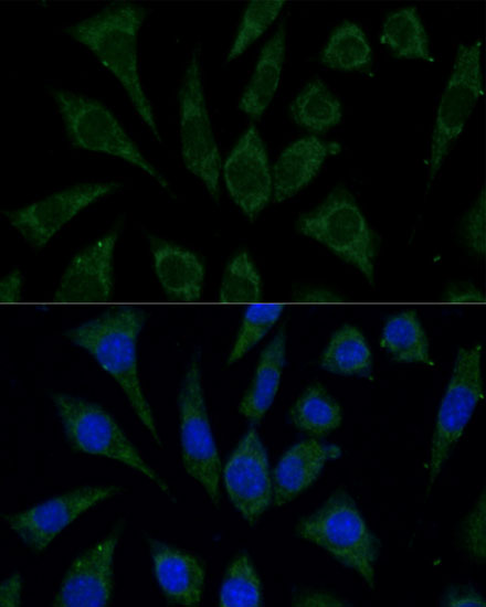
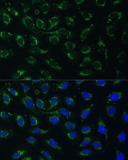
Immunofluorescence analysis of L929 cells using AKR1C2 Rabbit pAb (A1048) at dilution of 1:100 (40x lens). Secondary antibody: Cy3-conjugated Goat anti-Rabbit IgG (H+L) (AS007) at 1:500 dilution. Blue: DAPI for nuclear staining. |
|
|
Immunofluorescence analysis of U-2 OS cells using AKR1C2 Rabbit pAb (A1048) at dilution of 1:100 (40x lens). Secondary antibody: Cy3-conjugated Goat anti-Rabbit IgG (H+L) (AS007) at 1:500 dilution. Blue: DAPI for nuclear staining. |
Produktgarantie und fachkundiger Support
