
ATG7 Rabbit pAb, Unconjugated, Polyclonal
Artikelnummer:
ABB-A0691
- Bilder (8)


| Artikelname: | ATG7 Rabbit pAb, Unconjugated, Polyclonal |
| Artikelnummer: | ABB-A0691 |
| Hersteller Artikelnummer: | A0691 |
| Alternativnummer: | ABB-A0691-100UL,ABB-A0691-20UL,ABB-A0691-500UL,ABB-A0691-1000UL |
| Hersteller: | ABclonal |
| Wirt: | Rabbit |
| Kategorie: | Antikörper |
| Applikation: | ELISA, IF, IHC-P, WB |
| Spezies Reaktivität: | Human |
| Immunogen: | Recombinant protein (or fragment).This information is considered to be commercially sensitive. |
| Konjugation: | Unconjugated |
| Alternative Synonym: | GSA7, APG7L, SCAR31, APG7-LIKE, ATG7 |
| This gene encodes an E1-like activating enzyme that is essential for autophagy and cytoplasmic to vacuole transport. The encoded protein is also thought to modulate p53-dependent cell cycle pathways during prolonged metabolic stress. It has been associated with multiple functions, including axon membrane trafficking, axonal homeostasis, mitophagy, adipose differentiation, and hematopoietic stem cell maintenance. Alternative splicing results in multiple transcript variants. |
| Klonalität: | Polyclonal |
| Molekulargewicht: | 78 kDa |
| NCBI: | 10533 |
| UniProt: | O95352 |
| Reinheit: | Affinity purification |
| Sequenz: | LGFDTFVVMRHGLKKPKQQGAGDLCPNHPVASADLLGSSLFANIPGYKLGCYFCNDVVAPGDSTRDRTLDQQCTVSRPGLAVIAGALAVELMVSVLQHPEGGYAIASSSDDRMNEPPTSLGLVPHQIRGFLSRFDNVLPVSLAFDKCTACSSKVLDQYEREGFNFLAKVFNSSHSFL |
| Target-Kategorie: | ATG7 |
| Antibody Type: | Primary Antibody |
| Application Verdünnung: | WB,1:2000 - 1:8000|IHC-P,1:50 - 1:200|IF/ICC,1:50 - 1:200|ELISA,Recommended starting concentration is 1 µg/mL. Please optimize the concentration based on your specific assay requirements. |
| Anwendungsbeschreibung: | Cross-Reactivity: Human,Mouse,Rat. ResearchArea: Cancer,Signal Transduction,Cell Biology Developmental Biology,Apoptosis,Autophagy,Ubiquitin,Endocrine Metabolism,Mitochondrial metabolism,Immunology Inflammation,Cardiovascular,Heart. Shipping: Ice Bag |
|
|
Immunohistochemistry analysis of paraffin-embedded Human colon carcinoma using ATG7 Rabbit pAb (A0691) at dilution of 1:50 (40x lens). High pressure antigen retrieval performed with 0.01M Citrate buffer (pH 6.0) prior to IHC staining. |
|
|
Immunohistochemistry analysis of paraffin-embedded Human colon using ATG7 Rabbit pAb (A0691) at dilution of 1:50 (40x lens). High pressure antigen retrieval performed with 0.01M Citrate buffer (pH 6.0) prior to IHC staining. |
|
|
Western blot analysis of various lysates using ATG7 Rabbit pAb (A0691) at 1:2000 dilution incubated at room temperature for 1.5 hours. Secondary antibody: HRP-conjugated Goat anti-Rabbit IgG (H+L) (AS014) at 1:10000 dilution. Lysates/proteins: 25 µg per lane. Blocking buffer: 3% nonfat dry milk in TBST. Detection: ECL Basic Kit (RM00020). Exposure time: 10 s. |
|
|
Immunohistochemistry analysis of paraffin-embedded Mouse lung using ATG7 Rabbit pAb (A0691) at dilution of 1:50 (40x lens). High pressure antigen retrieval performed with 0.01M Citrate buffer (pH 6.0) prior to IHC staining. |
|
|
Immunohistochemistry analysis of paraffin-embedded Rat ovary using ATG7 Rabbit pAb (A0691) at dilution of 1:50 (40x lens). High pressure antigen retrieval performed with 0.01M Citrate buffer (pH 6.0) prior to IHC staining. |
|
|
Immunohistochemistry analysis of paraffin-embedded Rat spleen using ATG7 Rabbit pAb (A0691) at dilution of 1:50 (40x lens). High pressure antigen retrieval performed with 0.01M Citrate buffer (pH 6.0) prior to IHC staining. |
|
|
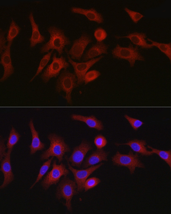
Immunofluorescence analysis of A-549 cells using ATG7 Rabbit pAb (A0691) at dilution of 1:50 (40x lens). Secondary antibody: Cy3-conjugated Goat anti-Rabbit IgG (H+L) (AS007) at 1:500 dilution. Blue: DAPI for nuclear staining. |
|
|
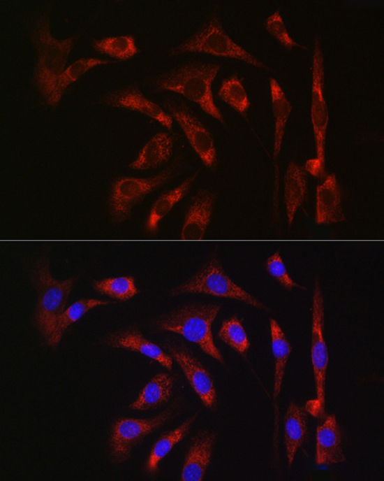
Immunofluorescence analysis of NIH/3T3 cells using ATG7 Rabbit pAb (A0691) at dilution of 1:50 (40x lens). Secondary antibody: Cy3-conjugated Goat anti-Rabbit IgG (H+L) (AS007) at 1:500 dilution. Blue: DAPI for nuclear staining. |
Produktgarantie und fachkundiger Support
