
FGF2 Rabbit pAb, Unconjugated, Polyclonal
Artikelnummer:
ABB-A0235
- Bilder (8)


| Artikelname: | FGF2 Rabbit pAb, Unconjugated, Polyclonal |
| Artikelnummer: | ABB-A0235 |
| Hersteller Artikelnummer: | A0235 |
| Alternativnummer: | ABB-A0235-100UL,ABB-A0235-20UL,ABB-A0235-1000UL,ABB-A0235-500UL |
| Hersteller: | ABclonal |
| Wirt: | Rabbit |
| Kategorie: | Antikörper |
| Applikation: | ELISA, IF, IHC-P, WB |
| Spezies Reaktivität: | Human |
| Immunogen: | Recombinant protein (or fragment).This information is considered to be commercially sensitive. |
| Konjugation: | Unconjugated |
| Alternative Synonym: | BFGF, FGFB, FGF-2, HBGF-2, FGF2 |
| The protein encoded by this gene is a member of the fibroblast growth factor (FGF) family. FGF family members bind heparin and possess broad mitogenic and angiogenic activities. This protein has been implicated in diverse biological processes, such as limb and nervous system development, wound healing, and tumor growth. The mRNA for this gene contains multiple polyadenylation sites, and is alternatively translated from non-AUG (CUG) and AUG initiation codons, resulting in five different isoforms with distinct properties. The CUG-initiated isoforms are localized in the nucleus and are responsible for the intracrine effect, whereas, the AUG-initiated form is mostly cytosolic and is responsible for the paracrine and autocrine effects of this FGF. |
| Klonalität: | Polyclonal |
| Molekulargewicht: | 31kDa |
| NCBI: | 2247 |
| UniProt: | P09038 |
| Reinheit: | Affinity purification |
| Sequenz: | PALPEDGGSGAFPPGHFKDPKRLYCKNGGFFLRIHPDGRVDGVREKSDPHIKLQLQAEERGVVSIKGVCANRYLAMKEDGRLLASKCVTDECFFFERLESNNYNTYRSRKYTSWYVALKRTGQYKLGSKTGPGQKAILFLPMSAKS |
| Target-Kategorie: | FGF2 |
| Antibody Type: | Primary Antibody |
| Application Verdünnung: | WB,1:500 - 1:1000|IHC-P,1:50 - 1:200|IF/ICC,1:50 - 1:200|ELISA,Recommended starting concentration is 1 µg/mL. Please optimize the concentration based on your specific assay requirements. |
| Anwendungsbeschreibung: | Cross-Reactivity: Human,Mouse,Rat. ResearchArea: Cancer,Invasion and Metastasis,Signal Transduction,Cell Biology Developmental Biology,Growth factors,Neuroscience,Stem Cells,Neural Stem Cells,Cardiovascular,Angiogenesis. Shipping: Ice Bag |
|
|
Western blot analysis of various lysates using FGF2 Rabbit pAb (A0235) at 1:2000 dilution. Secondary antibody: HRP-conjugated Goat anti-Rabbit IgG (H+L) (AS014) at 1:10000 dilution. Lysates/proteins: 25µg per lane. Blocking buffer: 3% nonfat dry milk in TBST. Detection: ECL Basic Kit (RM00020). Exposure time: 30s. |
|
|
Immunohistochemistry analysis of paraffin-embedded Mouse kidney using FGF2 Rabbit pAb (A0235) at dilution of 1:100 (40x lens). Microwave antigen retrieval performed with 0.01M PBS Buffer (pH 7.2) prior to IHC staining. |
|
|
Western blot analysis of lysates from 293T cells, using FGF2 Rabbit pAb (A0235) at 1:1000 dilution. Secondary antibody: HRP-conjugated Goat anti-Rabbit IgG (H+L) (AS014) at 1:10000 dilution. Lysates/proteins: 25µg per lane. Blocking buffer: 3% nonfat dry milk in TBST. Detection: ECL Enhanced Kit (RM00021). Exposure time: 180s. |
|
|
Immunohistochemistry analysis of paraffin-embedded Rat ovary using FGF2 Rabbit pAb (A0235) at dilution of 1:100 (40x lens). Microwave antigen retrieval performed with 0.01M PBS Buffer (pH 7.2) prior to IHC staining. |
|
|
Immunohistochemistry analysis of paraffin-embedded Human colon using FGF2 Rabbit pAb (A0235) at dilution of 1:100 (40x lens). Microwave antigen retrieval performed with 0.01M PBS Buffer (pH 7.2) prior to IHC staining. |
|
|
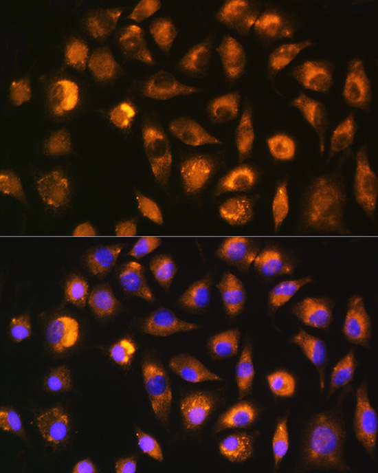
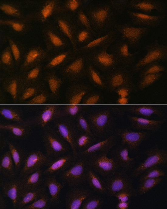
Immunofluorescence analysis of C6 cells using FGF2 Rabbit pAb (A0235) at dilution of 1:100 (40x lens). Secondary antibody: Cy3-conjugated Goat anti-Rabbit IgG (H+L) (AS007) at 1:500 dilution. Blue: DAPI for nuclear staining. |
|
|
Immunofluorescence analysis of L929 cells using FGF2 Rabbit pAb (A0235) at dilution of 1:100 (40x lens). Secondary antibody: Cy3-conjugated Goat anti-Rabbit IgG (H+L) (AS007) at 1:500 dilution. Blue: DAPI for nuclear staining. |
|
|
Immunofluorescence analysis of U-2 OS cells using FGF2 Rabbit pAb (A0235) at dilution of 1:100 (40x lens). Secondary antibody: Cy3-conjugated Goat anti-Rabbit IgG (H+L) (AS007) at 1:500 dilution. Blue: DAPI for nuclear staining. |
Produktgarantie und fachkundiger Support
