PD-1 Antibody is supplied in PBS containing 0.02% sodium azide and 50% glycerol.
Formulierung:
Liquid
Target-Kategorie:
PDCD1
Anwendungsbeschreibung:
Application Notes: PD-1 antibody can be used for detection of PD-1 by Western blot at 1 - 2 µg/mL. Antibody can also be used for immunohistochemistry starting at 5 µg/mL. For immunofluorescence start at 20 µg/mL. Flow cytometry at 0.1 µg/ml.Antibody validated: Immunohistochemistry in human samples, Immunocytochemistry in human samples, Immunofluorescence in human samples and Flow Cytometry in human samples. All other applications and species not yet tested
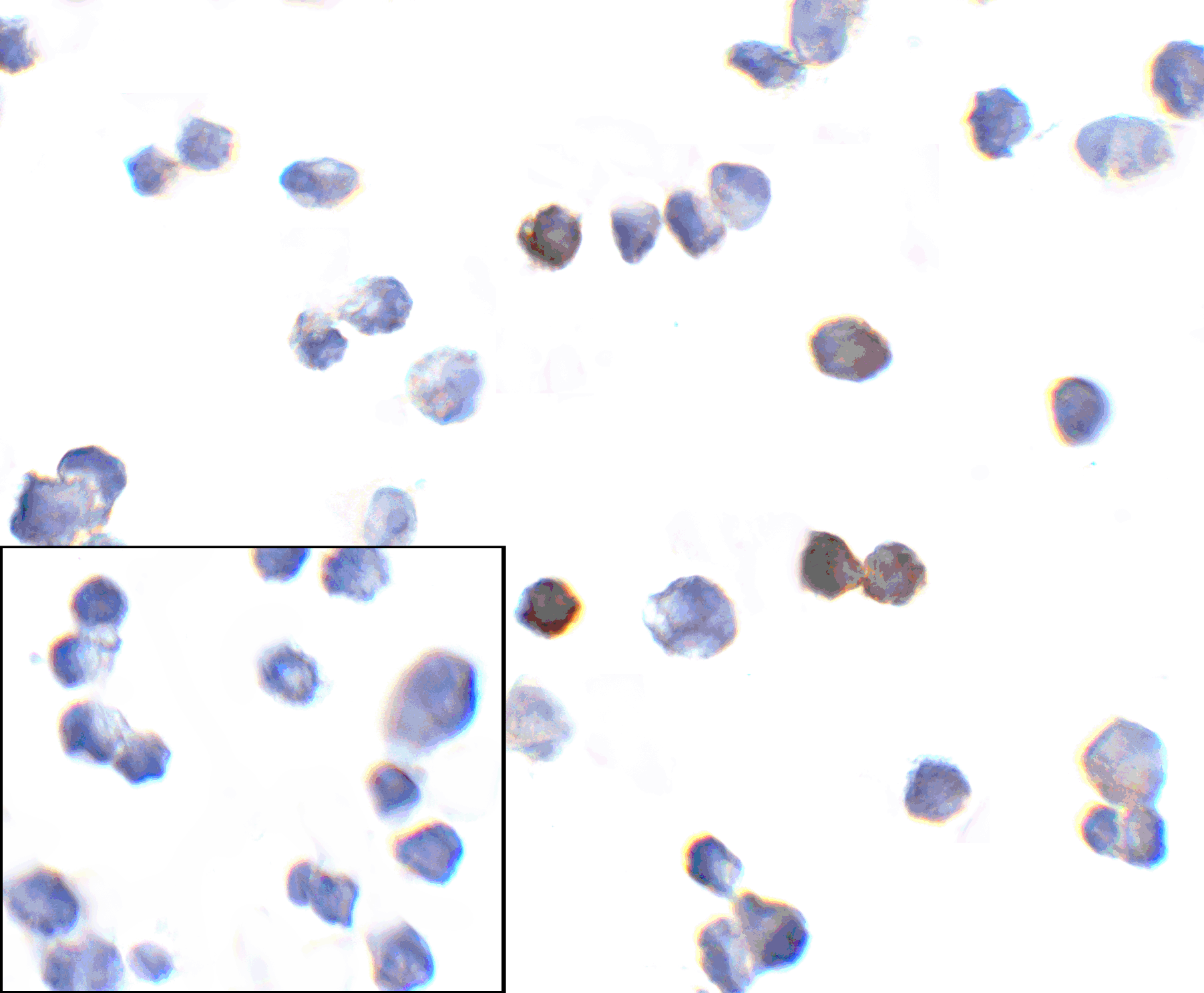
Immunocytochemistry of PD-1 in transfected 293 cells with PD-1 [10B3] antibody at 10 µg/mL. Lower left: Immunocytochemistry in transfected 293 cells with control mouse IgG antibody at 10 µg/mL.
(A) Immunohistochemistry of PD-1 in human spleen tissue with PD-1 [10B3] antibody at 10 µg/mL. (B) Immunohistochemistry in human spleen tissue with control mouse IgG staining at 10 µg/mL.
Immunofluorescence of PD-1 in human spleen tissue with PD-1 [10B3] antibody at 10 µg/mL. Green: PD1 Antibody [10B3] (orb1239734) Blue: DAPI staining
Flow cytometry analysis of PD-1 transfected 293 cells using PD-1 [10B3] antibody at 1 µg/ml. Blue: untransfected cells, Yellow: PD-1 transfected cells.
(A) Immunohistochemistry of PD-1 in human breast cancer tissue with PD-1 [10B3] antibody at 10 µg/mL. (B) Immunohistochemistry in human breast cancer tissue with control mouse IgG staining at 10 µg/mL.
Titration curve analysis of PD-1 mAbs to detect recombinant PD-1 in ELISA with orb1239745, orb1239769, orb1239763, orb1239738, and orb1239734 abs at decreasing concentrations.
* Mehrwertsteuer und Versandkosten nicht enthalten. Irrtümer und Preisänderungen vorbehalten